Molecular Mass Spectroscopy
250 likes | 528 Vues
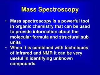
Molecular Mass Spectroscopy. Molecular structure Composition of mixtures Molecular mass spectra Ion Source Mass Spectrometers Applications. Molecular Mass Spectra. Removal of electron by electron bombardment In vapor phase Charged species is the molecular ion

Molecular Mass Spectroscopy
E N D
Presentation Transcript
Molecular Mass Spectroscopy • Molecular structure • Composition of mixtures • Molecular mass spectra • Ion Source • Mass Spectrometers • Applications
Molecular Mass Spectra • Removal of electron by electron bombardment • In vapor phase • Charged species is the molecular ion • Electron causes excitation and fragmentation • Major product is base peak • Assigned 100% relative abundance • Smaller fragments also form
Ion Sources • Ion source has profound effect on spectra • Gas phase source • Vaporized then ionized • Desorption source • Conversion of liquid or solid to gas • Hard source • Ion in excited state • Fragments produced • Soft source • Little fragmentation • Mainly ion of molecule
Electron-Impact Source • Sample as vapor • Ionized by beam of electrons • W or Re filament • 70 V potential • 1E-6 effective • M+e- ->M++2e- • High potentials in accelerating region • 1E3 to 1E4 volts • KE of ion in 1000 V • KE=qV=zeV • KE=1*1.6E-19 C*1000 V • 1.6E-16 J • KE independent of mass • Velocity varies with mass • KE=0.5mv2
Electron Impact Spectra • Energy from e- accelerated by 70V • Find in J/mol to compare bond energy • KE=eV • 1.6E-19 C *70 V=1.12E-17 CV/e- • For a mole • 1.12E-17 J*6.02E23 =6.7E6 J/mol • Bond energy 200 to 600 kJ/mol
Electron Impact Spectra • Sensitive method • Fragments useful in identification • Lack of molecular ion peak • M+, difficult to identify specie • Molecules must be in vapor phase • Stability issues in vapor phase • MW<1000 dalton
Isotopics • Isotopic variation can impact spectra
Chemical Ionization • Sample ionized by secondary ionization • Reagent gas ionized by electrons, then ionized reagent gas reacts with sample gas • Reagent to sample ratio • 1E3 to 1E4 • Methane most common reagent gas • CH4+ and CH3+ (about 90%), CH2+
Chemical Ionization • Produces ions that are 1 proton more or 1 proton less than molecule • Transfer of C2H5+ give M+29 peak • Field Ionization • Large electric field • 1E8 V/cm • Mainly produces M and M+1 peaks
Comparison of spectra • a-electron impact • b-field ionization • c-desorption
Large molecule desorption • Solid or liquid samples directly energized into gas phase • Molecular or protonated ion • Matrix Assisted Laser Desorption/Ionization (MALDI) • Soft Ionization • Sample dissolved in solution containing UV-absorber and solvent • Solution evaporated and precipitate formed • Pulsed laser used to excite precipitate • Molecular ion desorbed from surface of precipitate
Electrospray Ionization • Solution pumped through a needle • Needle is at kV potential compared to surrounding electrode • Droplets become charged • Solvent evaporates, droplets shrink and charge density increases • Can be combined with a number of methods • Useful for large molecules • M+, M2+
Fast Atom Bombardment • Samples in glycerol • Bombarded by Xe or Ar atoms • Several keV • Atoms and ions sputtered from surface • Production of fragments
Ion Trap Analyzer • Variable radio frequency voltage applied to the ring electrode • Ions of appropriate m/z circulate in stable orbit • scan radio frequency • heavier particles stable • lighter particles collide with ring electrode • ejected ions detected by transducer as an ion current
Applications • Identification of Pure Compounds: • Nominal M+ peak (one m/z resolution) (or (M+1)+ or (M-1)+) • gives MW (not EI) • Exact m/z (fractional m/z resolution) can give stoichiometry but not structure (double-focusing instrument) • Fragment peaks give evidence for functional groups • (M-15)+ peak methyl • (M-18)+ OH or water • (M-45)+ CO2H • series (M-14)+, (M-28)+, (M-42)+..sequential CH2 • loss in alkanes • Isotopic peaks can indicate presence of certain atoms • Cl, Br, S, Si • Isotopic ratios can suggest plausible molecules from M+, • Comparison with library spectra