Bibliography
Beta hemoglobin is a 147 amino acid protein: note that the final molecule is only 146 amino acids long. The first methionine is removed post-translationally. The 63rd amino acid in the final protein, histidine (H), is highlighted in red. In Hb M-Saskatoon, this amino acid is a tyrosine (Y).
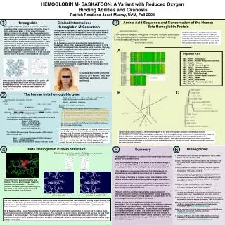

Bibliography
E N D
Presentation Transcript
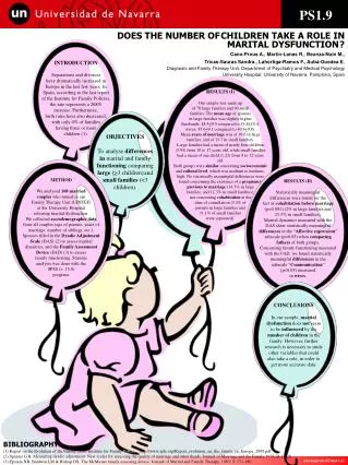
Beta hemoglobin is a 147 amino acid protein: note that the final molecule is only 146 amino acids long. The first methionine is removed post-translationally. The 63rd amino acid in the final protein, histidine (H), is highlighted in red. In Hb M-Saskatoon, this amino acid is a tyrosine (Y). Conserved histidine at position 63 in final protein Organism KEY HBB_PANTR _ Chimpanzee HBB_PANPA _ Pygmy chimpanzee (Bonobo) HBB_HUMAN _ Human HBB_GORGO _ Lowland gorilla HBB_HYLLA _Common gibbon HBB_SEMEN _ Hanuman langur HBB_ATEPA _ [Black spider monkey HBB_PITPI _ White-faced saki HBB_CEBAP _ Brown-capped capuchin HBB_RABIT _ Rabbit HBB_BALAC_Minke whale HBB_MACGG _ Australian ghost bat HBB_PIG _ Pig Conservation and Evolution A. Boxshade Diagram of the beta hemoglobin across 13 mammalian species (gi_4504349_ref_ is the human beta globin sequence). This is a highly conserved protein in mammals (see organism key). B. Drawtree - unrooted diagram showing the phylogeny relationships of beta hemoglobin protein. C. Drawgram- rooted phylogenic tree of beta hemoglobin protein. Both phylogenetic maps show that human Beta Hemoglobin is most like that of the Chimpanzee and the Bonobo our closest relatives. *Note: hemoglobin homologs are even found in bacteria! 1 3 Hemoglobin Clinical Information: Hemoglobin M-Saskatoon Amino Acid Sequence and Conservation of the Human Beta Hemoglobin Protein Hemoglobin (Hb) is the protein in red blood cells that reversibly binds oxygen and carries it from the lungs to all the cells of our body. It is the essential oxygen delivery protein in all animals. HbA, the form found in adult humans, is a tetrapeptide protein composed of two alpha chains (gene located on chromosome 16) and two beta chains (gene located on chromosome 11). Each peptide chain has one heme molecule bound to it. Each heme molecule has one atom of iron in the center of a protoporphyrin ring. The iron binds oxygen reversibly. To do so, iron must be in its reduced valence state (Fe+2). Methemoglobin (metHb) is the term used to describe hemoglobin in which the iron is in the oxidized valence state (Fe+3) and is unable to bind with oxygen. Hemoglobin M-Saskatoon is a beta peptide mutation and is one of seven known variants of hemoglobin in which the patient exhibits cyanosis (blue skin color) due to the presence of high levels of methemoglobin (metHb) in the red blood cells. For this reason the hemoglobin variants found in these patients are referred as “M” hemoglobins. Hb M-Saskatoon was first described in a Canadian family (in Saskatoon, CA) in 1956. Subsequently additional reports in 1978 and 1985 from Italy and India showed the same condition, implying the genetic change had occurred independently several times during human evolution. Hemoglobin M-Saskatoon has one allele where Histidine 63 is replaced by a Tyrosine (His63Tyr or H63Y). This leads to an accumulation of MetHb of up to 30 to 50%. MetHb has a blue/chocolate color which makes the patients skin look blue, called cyanosis.Inheritance patterns for Hb M variants are consistently heterozygous suggesting that the homozygous form is incompatible with life. removed from final protein 1 mvhltpeeks avtalwgkvn vdevggealg rllvvypwtq rffesfgdls tpdavmgnpk 61 vkahgkkvlg afsdglahld nlkgtfatls elhcdklhvd penfrllgnv lvcvlahhfg 121 keftppvqaa yqkvvagvan alahkyh HEMOGLOBIN M- SASKATOON: A Variant with Reduced Oxygen Binding Abilities and CyanosisPatrick Reed and Janet Murray, UVM, Fall 2008 63rd amino acid--histidine A Cyanosis due to the presence of up to 50% MetHb. Note blue skin hues especially on lips and neck. Heme molecule showing the iron atom at the center with bonding coordinates with the 4 nitrogen atoms of the protoporphyrin ring The iron atom is held within the heme molecule by two histidine amino acids of the peptide chain. B C The human beta hemoglobin gene 2 LOCUS NM_000518 626 bp mRNA linear PRI 12-OCT-2008 DEFINITION Homo sapiens hemoglobin, beta (HBB), mRNA. ACCESSION NM_000518 VERSION NM_000518.4 GI:28302128 chromosome: 11; Location: 11p15.5 NCBI data for a 626 bp mRNA transcript ORIGIN 1 acatttgctt ctgacacaac tgtgttcact agcaacctca aacagacacc atggtgcatc 61 tgactcctga ggagaagtct gccgttactg ccctgtgggg caaggtgaac gtggatgaag 121 ttggtggtga ggccctgggc aggctgctgg tggtctaccc ttggacccag aggttctttg 181 agtcctttgg ggatctgtcc actcctgatg ctgttatggg caaccctaag gtgaaggctc 241 atggcaagaa agtgctcggt gcctttagtg atggcctggc tcacctggac aacctcaagg 301 gcacctttgc cacactgagt gagctgcact gtgacaagct gcacgtggat cctgagaact 361 tcaggctcct gggcaacgtg ctggtctgtg tgctggccca tcactttggc aaagaattca 421 ccccaccagt gcaggctgcc tatcagaaag tggtggctgg tgtggctaat gccctggccc 481 acaagtatcactaagctcgc tttcttgctg tccaatttct attaaaggtt cctttgttcc 541 ctaagtccaa ctactaaact gggggatatt atgaagggcc ttgagcatct ggattctgcc 601 taataaaaaa catttatttt cattgc The human beta hemoglobin gene (HBB) consists of three exons and two introns and is 1606 bases. Exon 1, 1-142 (142 bp) Intron 1, 143-272 (130 bp) Exon 2 273-495 (223 bp) Intron 2, 496-1345 (850) Exon3 1346-1606 (261 bp) The mRNA is 626 bases long. Coding sequence (CDS) is from base 51 to base 494. Exon 1, 1-142 (includes 50 base 5’ UTR) (142 bases) Exon 2, 143-365 (223 bases) Exon3, 366-626 (Includes 132 base 3’ UTR) (261 bases) The leading UTR (black) is 50 bp long. The coding sequence starts at bp 51 (atg—in green).The 64th codon (bp 240-242: red) codes for a histidine (cat). A cytosine to thymine (c to t) change in the first base of the codon alters the code to a tyrosine (tat) seen in hemoglobin M-Saskatoon. The coding sequence of 441 bp (51 to 495—in orange) is for a 147 amino acid protein. The stop codon (taa—in green) starts at bp 492. The trailing UTR (black) starts at bp 495. The poly A signal sequence (red) begins at bp 602. 4 5 6 Beta Hemoglobin Protein Structure Bibliography Summary Substitution seen in Hemoglobin M-Saskatoon: a tyrosine for histidine at position 63 • Hematology: Clinical Principles and Applications, 3rd ed., Rodak, Fritsma and Doig, Saunders, 2007. • 2. Clinical Hematology: Principles, Procedures and Correlations, 2nd ed., Stienne-Martin, Lotspeich-Steininger and Koepke, Lippincott, 1998. • 3. Vella F, Kamuzora H, Lehmann H, Duncan B, Harold W, A second family with hemoglobin M Saskatoon in Saskatchewan, Clinical Biochemistry. 1974 Jun;7(2): pp186-91. • 4. Kedar PS, Nadkarni AH, Phanasgoankar S, Madkaikar M, Ghosh K, Gorakshakar AC, Colah RB, Mohanty D., Congenital methemoglobinemia caused by Hb-MRatnagiri (beta-63CAT-->TAT, His-->Tyr) in an Indian family. American Journal of Hematology. 2005: 79(2), pp. 168-70. • 5 .Da-Silva SS, Sajan IS, Underwood JP 3rd., Congenital methemoglobinemia: a rare cause of cyanosis in the newborn--a case report. Pediatrics., 2003: 112(2): pp158-61. • 6 .Stamatoyannopoulos G, Nute PE, De novo mutations producing unstable Hbs or Hbs M. II. Direct estimates of minimum nucleotide mutation rates in man. Human Genetics, 1982; 60(2): pp181-8. • 7. Hayashi A, Suzuki T, Fujita T, Diagnosis of Hb M disease by electron paramagnetic resonance spectra. Hemoglobin. 1980; 4(3-4): pp573-4. • 8. Multiple tools from National Center for Biotechnology Information (NCBI )U.S. National Library of Medicine 8600 Rockville Pike, Bethesda, MD 20894 http://www.ncbi.nlm.nih.gov/ • 9. Multiple tools from San Diego Supercomputer Center (SDSC) Biology WorkBench. University of California, San Diego. http://workbench.sdsc.edu/ • 10. Guex, N. and Peitsch, M.C. (1997) SWISS-MODEL and the Swiss-PdbViewer: An environment for comparative protein modeling. • Electrophoresis 18, 2714-2723. http://spdbv.vital-it.ch/ • Hemoglobin M-Saskatoon is a defect in the human beta hemoglobin protein that causes cyanosis. • The point mutation leading to this defect is a c to t base change at base 240 in the mRNA. This change leads to a codon change from c-a-t coding a histidine to t-a-t coding for tyrosine at amino acid 63. • The histidine is a conserved amino acid that interacts with the heme molecule and stabilizes the ferrous ion in reduced state. • The change of histidine to tyrosine results in oxidization of the ferrous ion and results in an inability to bind oxygen (MetHB form).. • This mutation is only seen in heterozygotes as the homozygote is most likely lethal. A heterozygote individual has up to 30 to 50% of beta hemoglobin in the MetHb form. • Patients with this condition have slightly higher hematocrits (measure of percent of RBCs in the blood) than normal suggesting that the decreased oxygen carrying capacity is compensated for by increasing the numbers of RBCs in the blood. • MetHb absorbs light at a different wave length than oxy-hemoglobin. This is normally evident in the color difference between arterial blood and venous blood. Arterial blood containing almost 100% oxy-hemoglobin is bright red. Venous blood, containing up to 75% deoxy-hemoglobin is blue. The permanent presence of high levels of MetHb in the capillary blood (close to the skin surface) in patients with Hb M-Saskatoon imparts a blue (cyanosis) color to the skin and is the most obvious phenoytpe of this rare mutation in the hemoglobin beta gene. Tyrosine Histidine View of the heme pocket formed by two helices. The iron atom (orange) has an O2 molecule (red) bound to it. The two histidine residues are shown stabilizing the iron atom in the center of the heme ring. The 63rd histidine is on the right. Wildtype human beta hemoglobin with histidine 63 Beta hemoglobin with tyrosine mutation at position 63 The 63rd histidine stabilizes the ferrous (Fe+2) atom in the heme ring and protects it from oxidation. During oxygen binding, the two helices of the heme pocket expands, permitting the entrance of an O2 molecule. Upon release of the O2 molecule, the heme pocket closes and the 63rd histidine moves closer to the iron atom and forms a bonding coordinate with it, protecting the reduced iron from oxidation. Hb M-Saskatoon has a weak Fe-peptide interaction due to a tyrosine for histidine substitution at position 63 of the beta chain, which leads to persistent oxidation of the iron molecule. The oxidized iron atom remains oxidized which renders the beta chain incapable of carrying oxygen. The image of beta hemoglobin with the tyrosine substitution at amino acid 63 shows negative interactions (in pink). Both tyrosine and histidine are polar amino acids. Histidine is positively charged while tyrosine is a larger neutral amino acid.