Rinse
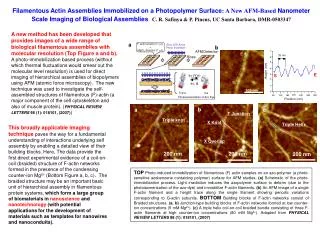
a. Blue LED Array Photo Irradiation. F-actin solution (1 m L) + MgCl 2 Solution (1 m L). AFM Detection. c. Rinse. Triple Helix. h n ( Vis). h n, D. 200 nm. Trans. Cis. Photoisomerization of Azo Dye. b. E. 7. 3 5 nm. 6. 5. b. a. 4. Height (nm). E. T Junction. S. 3.

Rinse
E N D
Presentation Transcript
a Blue LED Array Photo Irradiation F-actin solution (1 mL) + MgCl2 Solution (1 mL) AFMDetection c Rinse Triple Helix hn (Vis) hn, D 200 nm Trans Cis Photoisomerization of Azo Dye b E 7 35 nm 6 5 b a 4 Height (nm) E T Junction S 3 Triple knot 2 S X Knot 1 100 nm 0 200 160 80 120 240 280 320 Position (nm) X Overlap 200 nm 200 nm Filamentous Actin Assemblies Immobilized on a Photopolymer Surface: A New AFM-Based Nanometer Scale Imaging of Biological Assemblies C. R. Safinya & P. Pincus, UC Santa Barbara, DMR-0503347 A new method has been developed that provides images of a wide range of biological filamentous assemblies with molecular resolution (Top Figure a and b). A photo-immobilization based process (without which thermal fluctuations would smear out the molecular level resolution) is used for direct imaging of hierarchical assemblies of biopolymers using AFM (atomic force microscopy). The new technique was used to investigate the self-assembled structures of filamentous (F)-actin (a major component of the cell cytoskeleton and also of muscle protein). (PHYSICAL REVIEW LETTERS 98 (1): 018101, (2007)) This broadly applicable imaging technique paves the way for a fundamental understanding of interactions underlying self assembly by enabling a detailed view of their building blocks. Here, The data provide the first direct experimental evidence of a coil-on-coil (braided) structure of F-actin networks formed in the presence of the condensing counter-ion Mg2+ (Bottom Figure a, b, c). The braided structure may be an important basic unit of hierarchical assembly in filamentous protein systems, which form a large group of biomaterials innanoscienceandnanotechnology(with potential applications for the development of materials such as templates for nanowires and nanoconduits). TOP Photo-induced immobilization of filamentous (F) actin samples on an azo-polymer (a photo-sensitive azobenzenecontainingpolymer) surface for AFM studies. (a) Schematic of the photo-immobilization process. Light irradiation induces the azopolymer surface to deform (due to the photoisomerization of the azo-dye) and immobilize F-actin filaments. (b) An AFM image of a single F-actin filament and a height trace along the single filament showing periodic variations corresponding to G-actin subunits. BOTTOM Building blocks of F-actin networks consist of Braided structures.(a, b) Junction-type building blocks of F-actin networks formed at low counter-ion concentrations (5 mM Mg2+). (c)A triple helix coil-on-coil braided bundle consisting ofthree F-actin filaments at high counter-ion concentrations (80 mM Mg2+). Adapted from PHYSICAL REVIEW LETTERS 98 (1): 018101, (2007)
Phase Behavior and Interactions of Biomolecular MaterialsC. R. Safinya & P. Pincus, UC Santa Barbara, DMR- 0503347 Education: Multidisciplinary teams comprised of undergraduate and graduate students, and postdocs, with backgrounds inmaterials, physics, chemistry, and biology, are educated in methods to discover nature’s rules for assembling the molecular building blocks in distinct shapes and sizes for particular functions. The learned concepts enable development of advanced nanoscale materials for broad potential applications in electronic, chemical, and pharmaceutical industries. Outreach: Humphrey Kariuki(Top photo, right), a high school teacher in Oxnard, California, participated in the 2007 Materials Research Laboratory’s outreach RET (Research Experience for Teachers) Program. Together with his MentorMyung Chul Choi(bottom photo, middle, an exchange Postdoctoral Fellow in the PI’s lab from the Korean Advanced Institute of Science & Technology) they studied the binding properties of the protein tau to its substrate microtubule. Holger Hesse (Top photo, middle), an exchange undergraduate physics student from the Ludwigs Maximilians University (LMU) of Munich, was mentored by Joanna Deek(chemistry graduate student, Bottom photo, second from left), and Roy Beck(a Human Frontier Science Program Postdoctoral Fellow, top and bottom photos, left).The team formed an interdisciplinary working group studying the structure and phase behavior of Neurofilaments, which stabilize axon and dendrite neuronal processes. The structure work is conducted on the Materials Research Laboratory’s x-ray beam-lines (bottom photo, left), which are managed by Dr. Youli Liin collaboration with the PI’s group members. Kai Ewert(Project scientist & synthetic chemist, bottom photo, middle) and Alexandra Zidovska (Materials graduate student) mentored international exchange undergraduate physics student Heike Schirmer (Technical University of Munich), on the phase behavior of lipid-DNA complexes (Bottom photo, right).