Download
1 / 36
360 likes | 715 Vues
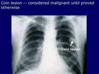
Coin lesion -- considered malignant until proved otherwise. Hamartoma - benign cartilaginous lesion. Cannon balls metastatic lesions. Metastases- irregularly sized and shaped lesions -- compare to previous CXR. Squamous cell carcinoma - large, irregular nuclei with keratin

E N D
Cannon balls metastatic lesions
Metastases- irregularly sized and shaped lesions -- compare to previous CXR
Squamous cell carcinoma - large, irregular nuclei with keratin more common men than women, most arise in bronchi, obstruction is presenting feature
Adenocarcinoma -- tall, columnar cells mucin (+) women, non-smokers <40 yoa, associated with scars, slow growing but metastasize early
Small cell carcinoma- basophilic cells with areas of necrosis, lymph-like cells (oat cells) derived from neuro-endocrine cells Associated with paraneoplastic syndromes
Endobronchial carcinoma This one is too high to be resected
A small endobronchial carcinoid (right). Histologically, there are well-defined nests of homogeneous cells with uniform nuclei (left).
Mesothelioma -- related to which type of asbestos? Amphibole (straight)
Pleural effusion must tap to determine if transudate or exudate
Squamous carcinoma -- hyperkeratosis, associated with alcohol and smoking Presentation: persistent hoarseness
Pneumocystis carinii hypoxemia, no infiltrate common in immunosuppressed pts
Lobar pneumonia -- classically what bt? Strep pneumo
Lobar pneumonia Stage 3 Gray hepatization -- PMNs ingesting and destroying RBCs
Acute bacterial pneumonia -- early red hepatization with neutrophils
Bronchopneumonia -- common organisms? Strep pneumo, H flu, Legionella, Pseudomonas
Bronchopneumonia -- some normal alveoli, congestion near bronchi
Interstitual pneumonia -- somealveoli filled, few inflammatory cells