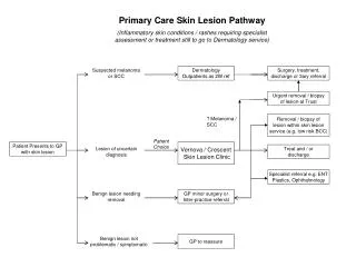
Skin Lesion
Skin Lesion. James Warneke, MD University of Arizona. Mr. Smith. Patient is a 64 year-old man with a history of a mole on his chest that has been present for years, but has recently grown is size. The mole has not bleed. History . What other points of the history do you want to know?.


Skin Lesion
E N D
Presentation Transcript
Skin Lesion James Warneke, MD University of Arizona
Mr. Smith • Patient is a 64 year-old man with a history of a mole on his chest that has been present for years, but has recently grown is size. The mole has not bleed.
History What other points of the history do you want to know?
History, Mr. Smith Consider the Following • Characterization of symptoms • Temporal sequence • Alleviating / Exacerbating factors: • Pertinent PMH, ROS, MEDS. • Relevant family hx. • Associated signs and symptoms
History, Mr Smith • Characteristics of mole • Change in color • Nodular areas • Family History of melanoma • History of sun exposure and sunburns • History of previous atypical moles
Differential DiagnosisBased on History • Dysplastic Nevus • Malignant Melanoma • Basal Cell Carcinoma • Squamous Cell Carcinoma • Junctional Nevus • Actinic Keratosis
Physical Examination What would you look for?
Physical Examination, Mr. Smith • General: fair skin and blue eyes • Skin: Multiple moles and evidence or solar skin damage on face and arms • Large mole on anterior chest with • Asymmetry, border irregularity, color variation and diameter greater than 6mm • Lymph nodes – none palpable in axilla, neck or groin
Biopsy of Lesion • Biopsy thickest area • Biopsy should be down to subcutaneous fat • Biopsy entire lesion if small • Large lesions should have punch biopsy or wedge • Orient extremity incisions in axial direction so re-excisions can be axial
Results of Biopsy • Melanoma What is important to check on the pathology report?
Biopsy Results • Pattern of Melanoma • Superficial spreading – most common • Nodular – vertical growth • Acral lentiginous – nails, palms and sole of foot, usually have in-situ precursor • Lentigo Maligna – in-situ melanoma in sun exposed area of the face and back of hand
Biopsy Results • Breslow’s Thickness – measured with an optic micrometer • Clark’s Level • I In-situ • II Papillary dermis • III Superficial reticular dermis • IV Deep reticular dermis • V Subcutaneous fat
Biopsy Results • Ulceration of Epithelium • Other Factors – deep margin involved with melanoma, regression, lymphocytic infiltration.
Biopsy Results of Mr. Smith • Superficial spreading melanoma with areas of nodular invasion • Breslow’s thickness 2.5mm • Clark’s level IV • Non-ulcerated • Deep margin free of melanoma
Laboratory and X-ray • What blood test should be ordered? • What X-ray studies are indicated?
Laboratory and X-ray • Serum LDH is indicated for lesions deeper than 1.0mm • PA and Lateral Chest X-ray for lesions deeper than 1.0mm
Laboratory and X-ray of Mr. Smith • LDH is within normal limits • CXR shows no evidence of metastatic disease
Further Management What should be done next?
Management Surgical Excision • How wide of an excision should be done? • When should a lymph node biopsy be planned?
Management of Mr. Smith • Margin of excision should be 2.0 cm from all borders of the pigmented lesion • Lesions <1.0mm 1.0 cm margins • Lesions 1.0-2.0 mm 1.0-2.0 cm margins • Lesions >2.0 mm 2.0 cm margins • The depth of the excision is to the underlying fascia
Management of Mr. Smith • For an acceptable cosmetic result, an ellipse of skin is usually excised with length 2.5-3.5 times the width. In this patient the lesion is wide, and the ellipse would be 6cm X 15cm • To close this defect primarily, the lateral edges are undermined for 2-3cm to allow the skin to stretch
Evaluation of Lymph Nodes • Lesions <1.0mm do not need lymph nodes biopsied • Lesions >1.0mm thickness should have sentinel lymph node biopsy • All lesions which have an enlarged palpable lymph node in an adjacent lymph node basin, should have that lymph node biopsied
Sentinel Lymph Node Biopsy • Lymphscintigraphy with Tc99 radiolabeled to colloid is done day of procedure to detect lymph drainage and to use intraoperatively with the gamma probe • Lymphazurin blue dye is injected into the dermis next to the melanoma to visually detect the sentinel lymph node
Sentinel Lymph Nodes in Mr. Smith • Sentinel lymph node biopsy found two lymph nodes which where blue • Both blue lymph nodes were hot with the hand-held gamma probe • Pathology by routine histology and immunohistochemistry did not detect any melanoma in the lymph nodes
Staging of Mr. Smith’s Melanoma • Primary Tumor (T) 2.5mm with no • ulceration is T3a • Regional Lymph Nodes (N) no regional node metastasis is NO • Metastasis (M) none is MO • Stage is IIAT3a NO MO
Prognosis • What is the estimated 10 year survival of Mr. Smith?
10 Year Survival of Mr. Smith • Melanoma T3a with NO has a 10 year survival of 65% • With the inclusion of sentinel lymph node biopsy, the micrometastatic nodes with melanoma will have a worse prognosis of 50%, and the negative sentinel nodes will have a increased survival to 80-90 %
Summary of Melanoma • 54,200 new melanomas per year in US • 7600 deaths from melanoma per year in US • 1 in 57 white males • 1 in 81 white females • 89% 5-year survival for 1992-1998
Acknowledgment The preceding educational materials were made available through theASSOCIATION FOR SURGICAL EDUCATION In order to improve our educational materials wewelcome your comments/ suggestions at: feedbackPPTM@surgicaleducation.com