Download

1 / 19
210 likes | 488 Vues
The Skin lesion. Kenny DeSart , MD. University of Florida Oral Exam Review. Introduction. Main goal is to differentiate benign vs. malignant lesions

E N D
The Skin lesion Kenny DeSart, MD University of Florida Oral Exam Review
Introduction Main goal is to differentiate benign vs. malignant lesions DDx benign: congenital nevus, acquired nevus, atypical nevus, melanosis of the genitalia, blue nevus, solar lentigo, hemanioma, Kaposi sarcoma, pyogenic granuloma, dermatofibroma, angiokeratoma DDx malignant: basal cell carcinoma, squamous cell carcinoma, melanoma
History • Assess melanoma risk factors • Family/Personal history of melanoma • Fair skin • History of sun exposure • Presence of actinic keratoses • History of the lesion • Change in size, shape, color, itching, bleeding
Physical examination • Multiple benign nevi higher risk of melanoma • ABCDE • Asymmetry • Irregular Borders • Variegated (mixed) Color • Large Diameter (>6mm) • Elevated surface • In-transit metastases • Palpate lymph nodes Skinipedia.org
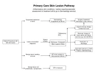
Workup/Treatment • <2cm lesion: Excisional biopsy unless cosmetically sensitive area • Full thickness ellipse, 1-4mm margin normal skin • Length of ellipse should be ~3x the width to allow tension-free closure • No electrocautery, no shave biopsies • >2cm lesion or cosmetically sensitive area: Incisional or punch bx
Depth of Invasion • Clark’s level: epidermis, papillary dermis, reticular dermis, and subcutaneous fat • Limited use (Clark’s IV or V and <1mm depth) • Breslow Depth: tumor thickness from the epidermal surface to the deepest point of the tumor's extension into tissue • Used in American Joint Committee of Cancer (AJCC) TNM classification
Surgical Treatment • Wide local excision (WLE) is the cornerstone of treatment • Margin of excision depends on depth • <1mm: 1cm margin • 1-4mm: 2cm margin • >4mm: 2-3cm margin • Primary closure or skin graft • Resect all clinically positive nodes • Sentinel lymph node biopsy • Not needed if <1mm (WLE 95% cure • Unless Clark’s IV or V • Perform in >1mm and no clinically evident nodes (T1)
Surgical Treatment • Positive regional lymph nodes – consider radiation therapy • Can resect isolated metastases (lung/liver) • In-transit metastases – found along lymphatic drainage between primary lesion and regional lymph nodes • Tx: WLE and lymph node dissection • Tx: palliative radiation if unresectable • Tx: isolated limb perfusion (usu. leg) Unboundmedicine.com
Labs and Imaging • Staging and workup • <1mm: no labs/imaging • More advanced disease: • Serum lactose dehydrogenase (LDH) level • Liver function tests (Liver #2 metastasis) • Chest X-ray (Lung #1 most common metastasis) • Body CT/Brain MRI to eval for distant mets
TNM Classification • Tumor – a (no ulceration), b (ulceration) • T1 ≤ 1 mm • T2 1.01-2 mm • T3 2.01-4 mm • T4 >4 mm • Node • N1 – 1 node • N2 – 2-3 nodes • N3 – 4+ nodes • Metastasis • M1a – distant skin, subq, or lymph node and normal LDH (lactate dehydrogenase) • M1b – lung metastases with normal LDH • M1c – Visceral metastases with normal LDH • OR any distant metastases with elevated LDH
Types of Melanomas • Superficial spreading – most common (70%) • Arises from pre-existing nevi • Nodular – aggressive (15-30%) • Mostly vertical growth (no radial growth) • Can rarely lack pigment • High metastatic rate • Lentigomaligna – older patients (4-10%) • Typical over face, typically large (>3cm) • Arises from macular brown nevus • Less aggressive, best prognosis • Acrallentiginous – palms or soles in patients with dark skin (2-8%) • Most aggressive • Highest metastatic rate • Ocular melanoma – most common non-cutaneous melanoma
Superficial spreading Sabiston 18th edition
Nodular Sabiston 18th edition
Lentigomaligna Sabiston 18th edition
Acrallentiginous Sabiston 18th edition
Melanoma Pearls • Prognosis • Tumor thickness is most important • Ulceration also important • Distant metastases • Lung > liver > brain > bone • Survival is 11mo for lung, 2-6 mo for others • Solitary brain metastasis should be resected • Can bleed, some can live 5+ years with resection and radiation therapy • The most common type of metastasis to small bowel is melanoma
Basal Cell Carcinoma Most common malignancy in US Pearly appearance, rolled borders Locally invasive, rare mets 3-5mm margin MOHs surgery
Squamous Cell Carcinoma Actinic keratosis is precursor lesion Can arise is chronic wounds/scars/burn scar 0.5-2cm margins MOHs surgery Regional lymphadenectomy for clinically positive nodes