Chapter 5: Cell Division
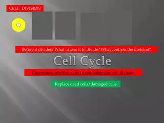
Chapter 5: Cell Division. Cell Increase and Decrease. Cell division increases the number of somatic (body) cells , and consists of: Mitosis (division of nucleus) Cytokinesis (division of cytoplasm) Apoptosis (cell death) decreases the number of cells. The cell cycle. Fig. 5.1.

Chapter 5: Cell Division
E N D
Presentation Transcript
Cell Increase and Decrease • Cell division increases the number of somatic (body) cells, and consists of: • Mitosis (division of nucleus) • Cytokinesis (division of cytoplasm) • Apoptosis (cell death) decreases the number of cells.
The cell cycle Fig. 5.1 Most of the cell cycle is spent in interphase. Following interphase, the mitotic stage of cell division occurs.
The stages of interphase • G1 stage – cell growth, # of organelles doubles • S stage – DNA synthesis and replication occurs • G2 stage – protein synthesis for cell division
The cell cycle Fig. 5.1 Following interphase is the M stage, including mitosis and cytokinesis. The cell cycle ends when cytokinesis, the cleaving of the cytoplasm, is complete. Interphase Mitosis and cytokinesis The Cell Cycle: G1 S G2 M
Fig. 5.1 G1 checkpoint M checkpoint G2 checkpoint The cell cycle is controlled at three checkpoints. DNA damage can stop the cell cycle at the G1 and G2 checkpoint. If chromosomes are not properly aligned, cell cycle stops at the M stage
Apoptosis • Apoptosis - programmed cell death. • - occurs because of two sets of enzymes called capsases. • “initiators” - receive a signal to activate the “executioners”. • Executioners activate enzymes that tear apart the cell and its DNA.
Maintaining the Chromosome Number • During interphase, the DNA and associated proteins is are called chromatin. • During Mitosis , the chromatin condenses to form highly compacted structures called chromosomes.
Overview of Mitosis • The haploid (n) number of chromosomes = the number of kinds of chromosome. • The diploid (2n) number of chromosomes = two chromosomes of each kind. • Humans have 23 types (haploid) of chromosomes, so we have a total pf 46 chromosomes (diploid).
DNA replication takes place before nuclear division occurs. Page 85 A duplicated chromosome is made of two sister chromatids held together at the centromere. During mitosis, the centromeres divide and the sister chromatids become daughter chromosomes. genetically identical.
Mitosis overview Fig. 5.3 Diploid
Following mitosis, a diploid parental cell gives rise to two diploid daughter cells, or 2n → 2n. Mitosis occurs when tissues grow (throughout the lifespan of the organism) or when repair occurs.
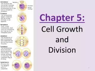
Mitosis in Detail • Mitosis has four phases: prophase, metaphase, anaphase, and telophase.
Late Interphase Fig. 5.4 Chromatin is condensing into chromosomes. Centrosomes have duplicated.
Early Prophase Fig. 5.5 Duplicated chromosomes are visible. Nuclear envelope is fragmenting and nucleolus will disappear. Spindle fibers appear between the separating centrosomes.
Late Prophase Fig. 5.5 Spindle is in process of forming Centromeres of chromosomes are attaching to centromeric spindle fibers Chromosomes have no particular orientation
Metaphase Fig. 5.5 Spindle is fully formed (poles, asters, and fibers) Chromosomes are at the metaphase plate of the fully formed spindle Metaphase plate
Anaphase Fig. 5.5 Centromeres divide Sister chromatids separate Daughter chromosomes begin to move toward the opposite poles of the spindle
Telophase Fig. 5.5 Spindle disappears Nuclear envelope reappears Chromosomes become diffuse chromatin again Cleavage furrow visible
How Plant Cells Divide • Plant cells lack centrioles and asters, but have a centrosome and spindle and the same four stages of mitosis.
Cytokinesis in Plant and Animal Cells • Cytokinesis, or cytoplasmic cleavage, accompanies mitosis. • Cleavage of the cytoplasm begins in anaphase, but is not completed until just before the next interphase.
Cytokinesis in Plant Cells • The rigid cell wall surrounding plant cells does not permit cytokinesis by furrowing. • The Golgi apparatus releases vesicles that microtubles move to the cell plate forming between the two new cells. • New plant cell walls form and are later strengthened by cellulose fibers.
Cytokinesis in plant cells Fig. 5.7
Cytokinesis in Animal Cells Fig. 5.8 In animal cells, a cleavage furrow begins at the end of anaphase. A contractile ring (actin and myosin) filaments slowly forms a constriction between the two daughter cells.
Cell Division in Prokaryotes • The process of asexual reproduction in prokaryotes is called binary fission. • The two daughter cells are identical to the original parent cell, each with a single chromosome. • Following DNA replication, the two resulting chromosomes separate as the cell elongates.
Reducing the Chromosome Number • Meiosis reduces the chromosome number such that each daughter cell has only one of each kind of chromosome (Reduction Division). • Meiosis ensures that the next generation will have: • the diploid number of chromosomes • A single copy of each type of chromosome from each parent.
Overview of meiosis Fig. 5.9
Meiosis in Detail • The same four phases seen in mitosis – prophase, metaphase, anaphase, and telophase – occur during meiosis I and meiosis II. • Interkinesis - period between meiosis I and meiosis II. • No replication of DNA occurs during interkinesis because the DNA is already duplicated.
Meiosis I in an animal cell From Fig. 5.12 Crossing over can occur here Homologous chromosomes line up
From Fig. 5.12 Haploid – but chromosomes are duplicated
From Fig. 5.13 Meiosis II No need for duplication – just separation Haploid Metaphase II Anaphase II Prophase II Telophase II
Comparison of Meiosis with Mitosis • In both mitosis and meiosis, DNA replication occurs only once during interphase. • Mitosis requires one division while meiosis requires two divisions. • Two diploid daughter cells result from mitosis; four haploid daughter cells result from meiosis.
Genetic Recombination • There are two sources of genetic recombination during meiosis: • crossing-over of nonsister chromatids and • independent assortment of homologous chromosomes. • Both events assure new genetic combinations in the offspring. • Genetic recombination is the process by which the combination of genes in an organism's offspring becomes different from the combination of genes in that organism
Synapsis and Crossing-over Occurs During Prophase I Fig. 5.10
1 M 1 M 1 F 1 F 1 M 1 F 1 F 1 M 3 M 3 F 3 M 3 F 3 F 3 M 3 M 3 F 2 F 2 F 2 M 2 M 2 M 2 F 2 M 2 F Independent assortment Fig. 5.11 Organism with 3 types of chromosomes. - 3 types of chromosomes, 2 copies of each. - one copy from each parent. Large = chromosome 1 Medium = chromosome 2 Small = chromosome 3
oogenesis spermatogenesis Life cycle of humans The human life cycle requires both mitosis and meiosis.
Sources of Genetic Recombination in Humans • Independent assortment of chromosomes during metaphase I • Crossing-over during prophase I • Upon fertilization, recombination of chromosomes occurs.