Cell division
Cell division. Plant physiology. CELL CYCLE. Cell division is a very important process in all living organisms. During the division of a cell, DNA replication and cell growth also take place.

Cell division
E N D
Presentation Transcript
Cell division Plant physiology
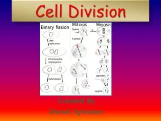
CELL CYCLE • Cell division is a very important process in all living organisms. • During the division of a cell, DNA replication and cell growth also take place. • All these processes, i.e., cell division, DNA replication, and cell growth, hence, have to take place in a coordinated way to ensure correct division and formation of progeny cells containing intact genomes. • The sequence of events by which a cell duplicates its genome, synthesises of the other constituents of the cell and eventually divides into two daughter cells is termed cell cycle.
Phases of Cell Cycle • The cell cycle is divided into two basic phases: • Interphase • M Phase (Mitosis phase
M phase • The M Phase represents the phase when the actual cell division or mitosis occurs . • The M Phase starts with the nuclear division, corresponding to the separation of daughter chromosomes (karyokinesis) and usually ends with division of cytoplasm (cytokinesis).
Interphase • The interphase represents the phase between two successive M phases. • The interphase, though called the resting phase, is the time during which the cell is preparing for division by undergoing both cell growth and DNA replication in an orderly manner. • The interphase is divided into three further phases: • G1 phase (Gap 1) • S phase (Synthesis) • G2 phase (Gap 2)
Interphase • G1 phase - corresponds to the interval between mitosis and initiation of DNA replication. During G1 phase the cell is metabolically active and continuously grows but does not replicate its DNA. • S or synthesis phase - marks the period during which DNA synthesis or replication takes place. During this time the amount of DNA per cell doubles. However, there is no increase in the chromosome number. In animal cells, during the S phase, DNA replication begins in the nucleus, and the centriole duplicates in the cytoplasm.
G2 phase - proteins are synthesised in preparation for mitosis while cell growth continues. G0 phase - Some cells in the adult animals do not appear to exhibit division (e.g., heart cells) and many other cells divide only occasionally, as needed to replace cells that have been lost because of injury or cell death. These cells that do not divide further exit G1 phase to enter an inactive stage called quiescent stage (G0) of the cell cycle. Cells in this stage remain metabolically active but no longer proliferate unless called on to do so depending on the requirement of the organism.
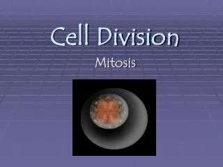
M-Phase • Four Phases • Prophase • Metaphase • Anaphase • Telophase
Prophase • Prophase is marked by the initiation of condensation of chromosomal material. • The chromosomal material becomes untangled during the process of chromatin condensation. • The centriole, which had undergone duplication during S phase of interphase, now begins to move towards opposite poles of the cell.
M -Phase • Prophase
Metaphase • The complete disintegration of the nuclear envelope marks the start of the second phase of mitosis • the chromosomes are spread through the cytoplasm of the cell. • By this stage, condensation of chromosomes is completed and they can be observed clearly under the microscope. • This then, is the stage at which morphology of chromosomes is most easily studied. • At this stage, metaphase chromosome is made up of two sister chromatids, which are held together by the centromere • These structures serve as the sites of attachment of spindle fibres (formed by the spindle fibres) to the chromosomes that are moved into position at the centre of the cell.
Metaphase…………. • Hence, the metaphase is characterised by all the chromosomes coming to lie at the equator with one chromatid of each chromosome connected by its kinetochore to spindle fibres from one pole and its sister chromatid connected by itskinetochore to spindle fibres from the opposite pole. • The plane of alignment of the chromosomes at metaphase is referred to as the metaphase plate. • The key features of metaphase are: • Spindle fibres attach to kinetochores of chromosomes. • Chromosomes are moved to spindle equator and get aligned along metaphase plate through spindle fibres to both poles.
Anaphase • At the onset of anaphase, each chromosome arranged at the metaphase plate is split simultaneously • and the two daughter chromatids begin their migration towards the two opposite poles. • As each chromosome moves away from the equatorial plate, • the centromere of each chromosome is towards the pole.
Anaphase….. • anaphase stage is characterised by the following key events: • Centromeres split and chromatids separate. • Chromatids move to opposite poles.
Telophase • At the beginning of the final stage of mitosis, i.e., telophase, the chromosomes that have reached their respective poles decondense and lose their individuality. • The individual chromosomes can no longer be seen • chromatin material tends to collect in a mass in the two poles
Telophase…….. • This is the stage which shows the following key events: • Chromosomes cluster at opposite spindle poles and their identity is lost as discrete elements. • Nuclear envelope assembles around the chromosome clusters. • Nucleolus, golgi complex and ER reform.
Cytokinesis • Mitosis accomplishes not only the segregation of duplicated chromosomes into daughter nuclei (karyokinesis), • but the cell itself is divided into two daughter cells by a separate process called cytokinesis at the end of which cell division is complete.
In an animal cell, this is achieved by the appearance of a furrow in the plasma membrane. • The furrow gradually deepens and ultimately joins in the centre dividing the cell cytoplasm into two.
Plant cells however, are enclosed by a relatively inextensible cell wall, therefore they undergo cytokinesis by a different mechanism. • In plant cells, wall formation starts in the centre of the cell and grows outward to meet the existing lateral walls.
The formation of the new cell wall begins with the formation of a simple precursor, called the cell-plate that represents the middle lamella between the walls of two adjacent cells. • At the time of cytoplasmic division, organelles like mitochondria and plastids get distributed between the two daughter cells.
Significance of Mitosis • Mitosis results in the production of diploid daughter cells with identical genetic complement usually. • The growth of multicellular organisms is due to mitosis. • A very significant contribution of mitosis is cell repair. • The cells of the upper layer of the epidermis, cells of the lining of the gut, and blood cells are being constantly replaced. • Mitotic divisions in the meristematic tissues – the apical and the lateral cambium, result in a continuous growth of plants throughout their life.