Download
1 / 17
170 likes | 344 Vues
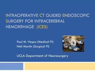
I ntraoperative C T guided E ndoscopic S urgery for Intracerebral Hemorrhage (ICES). Paul M. Vespa, MD Neil Martin, MD Jennifer Varma, RN, NP UCLA ICES Surgical Center Dan Hanley,MD PI MISTIE/ICES Coordinating Center Johns Hopkins. Experimental Procedure.

E N D
Intraoperative CT guided Endoscopic Surgery for Intracerebral Hemorrhage (ICES) Paul M. Vespa, MD Neil Martin, MD Jennifer Varma, RN, NP UCLA ICES Surgical Center Dan Hanley,MD PI MISTIE/ICES Coordinating Center Johns Hopkins
Experimental Procedure • Initial Screen, Estimation of clot size (cc) • Prepare for OR, plan trajectory, • Call Dr. Martin • Stereotactic placement of burr hole • Stereoactic trajectory for endoscope • Suction of hematoma (start at 50 mm Hg) • Measurement of extracted clot volume (cc) • Hemostasis • Placement of hematoma drain • Repeat imaging • CT Post-op, CT Days 1-4, MRI day 7
Registry case (05-S06) • 75 year-old female • History of meningioma, s/p resection and radiation therapy in 1999 • Restrained driver involved in MVA, noted to be driving erratically before hitting another vehicle • Upon examination by EMS, found to have left-sided arm and leg weakness • Initial head CT demonstrated large right-sided hematoma, estimated at about 50 cc
Registry Case • Screened for ICES, determined to be a good candidate • Informed consent obtained • CTA performed and did not show evidence of an aneurysm or vascular malformation • Stability CT showed no further extension of the hematoma • Patient to OR approximately < 12 hours following initial CT
Operative Procedure • Right parietal burr hole • Initial depth 9 cm, suctioned up to 300 mm Hg, no clot obtained • Repositioned endoscope to a depth of 7 cm, suctioned up to 300 mm Hg and 32 cc of clot obtained; about a 60% clot reduction • Bleeding vessels controlled with monopolar and DDAVP infusion • Hematoma catheter left in place
Post-op BrainLAB screen shots:(yellow – preop, orange – postop)
Key Points • Surgical Planning with Dr. Martin • Steps in protocol with documentation in OR • Database documentation of ICU days • Serial Imaging: CT daily x 4 days, MRI at day 7 • Submit registry cases for review to the coordinating center and surgical center