Download
1 / 13
150 likes | 408 Vues
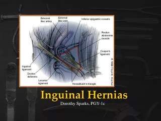
Complications of Hiatal Hernias. Krista Fajman, M.D. September 23, 2009. Hiatal Hernias. Herniation of organs of the abdomen into the thoracic cavity through the esophageal hiatus at the diaphragm

E N D
Complications of Hiatal Hernias Krista Fajman, M.D. September 23, 2009
Hiatal Hernias • Herniation of organs of the abdomen into the thoracic cavity through the esophageal hiatus at the diaphragm • Weakening of the diaphragmatic hiatus thought to be caused by repetitive stress of swallowing, abdominal straining, vomiting in addition to contraction of the esophageal longitudinal muscle induced by reflux and acidification of the mucosa.
Classification • Type I (Sliding Hiatal Hernia) • > 95% of cases, increasing incidence with age • Hiatus widens and phrenoesophageal membrane loosens gastroesophageal junction and fundus of the stomach displaced above the diaphragm. • Hernia contained within the posterior mediastinum. • Diagnose with barium swallow or endoscopy if > 2 cm. Esophageal manometry can diagnose smaller hernias.
Classification continued • Types II, III, IV (Paraesophageal Hernia) • Type II – GE junction fixed. Progressive enlargement of the hiatus • Type III (Mixed Hernia) – Enlargement of the hiatal hernia, displacement of the lower esophageal sphincter and gastric fundus above the diaphragm • Type IV – Associated with herniation of the colon, spleen, pancreas, and small intestine • 5-10% of hernias. • Unclear etiology but associated with weakening of the gastrosplenic and gastrocolic ligaments. • Best diagnosed with barium swallow although can usually be seen on endoscopy. CT or MRI helpful if you suspect herniation of organs other than the stomach.
Symptoms • May be asymptomatic • Reflux • Chest, epigastric or substernal pain • Dysphagia • Postprandial fullness • Nausea, vomiting • Gastritis • Chronic blood loss from gastric ulceration or erosions within the hernia • Respiratory complications
Hiatal Hernia Treatment • Repair of asymptomatic type I hernias rarely require surgical repair. If pt has GERD, medical or surgical treatment is indicated to control the reflux. • Types II, III, and IV – Even if asymptomatic, imposes a certain risk on the patient. • Never regress although may enlarge. • Can result in herniation of the stomach or other abdominal organs into the thorax. • If untreated can pose a risk of serious complications. • Nonoperative mortality rate = as high as 80% • Mortality rate from acute gastric volvulus = 15-20% • Mortality rate from chronic gastric volvulus ranges up to 13% • Once identified treat surgically.
Gastroesophageal Reflux • Estimated prevalence of 14-20% in the general population • Diagnose with endoscopy rule out Barrett’s, adenocarcinoma, ulcers. Also consider H. pylori, manometry • First line treatment – Lifestyle modification, Antacids, H2 blockers and PPIs • Complications of chronic PPI use: • Increased risk of hip fracture in pts > 50 years old • Increased risk of infectious gastroenteritis • Increased risk of C. difficile colitis • Decreased effectiveness of Plavix
Treatment for refractory GERD associated with hernias • Consider TCAs in those that do not respond • Fundoplication - the proximal stomach is wrapped around the LES increased LES pressure. • Surgical risks include frequent need for revision, dysphagia, flatulence, difficulty belching, bloating, abdominal pain, diarrhea, constipation. • No difference in rates of Barrett’s metaplasia or adenocarcinoma between medical or surgical management.
Gastric Volvulus • Uncommon, although most common complication of paraesophageal hernias. • Paraesophageal hernias progressively enlarge eventually the stomach herniates through the hiatus forming an upside down stomach in the thoracic cavity. • Associated with dysphagia or postprandial pain. • Twisting of up to 180 degrees may cause no obstruction
Gastric Volvulus • Organoaxial • Most common • Associated with diaphragmatic defects • Vascular compromise common • Mesenteric Axis • Chronic symptoms more common • Diaphragmatic defect less common
References • Ho, M., Maddox, T., Wang, L., Fihn, S., Jesse, R., Peterson, E., Rumsfeld, J. “Risk of Adverse Outcomes Associated with Concomitant Use of Clopidogrel and Proton Pump Inhibitors Following Acute Coronary Syndrome.” JAMA. March 4, 2009. 301: 937-944. • Kahrilas, P., Talley, N., and Bonis, P. “Hiatus Hernia” UpToDate Online. December 15, 2008 • Kahrilas, P. “Gastroesophageal Reflux Disease.” New England Journal of Medicine. Vol 359: 1700-1707. October 16, 2008. • Kasper, Braunwald, Fauci, Hauser, Longo and Jameson. Harrison’s Principles of Internal Medicine, 16th Edition. • LearningRadiology.com • Sivaskarin, S., Kawamura, A., Lombardi, D., Nesto, R. “Gastric Volvulus Presenting as an Acute Coronary Syndrome.” Texas Heart Institute Journal. 2006; 33(2): 266-268.