HERNIAS
HERNIAS. Dr David Swar General Surgery Qilu Hospital Shandong University. Definition. A hernia is the protrusion of an organ through its containing wall. Composition of a hernia. The sac The covering of the sac The content of the sac. Composition of a hernia. The sac :

HERNIAS
E N D
Presentation Transcript
HERNIAS Dr David Swar General Surgery Qilu Hospital Shandong University
Definition • A hernia is the protrusion of an organ through its containing wall.
Composition of a hernia • The sac • The covering of the sac • The content of the sac
Composition of a hernia • The sac : • It is a diverticulum of peritoneum and is made up of three parts : • The mouth, • The neck and • The body of the sac.
Composition of a hernia • The covering: • Coverings are derived from the layers of abdominal wall through which the sac pass • Contents: • can be • Omentum = omentocle • Intestine = enterocele • Portion of circumference of intestine = Richter’s hernia • Portion of the bladder • Ovary(with or without oviduct) • Meckel’s diverteculum =Littre’s hernia
Etiology • Hernias occur at sites of weakness in the wall • This weakness may be : • Normal (physiological) weakness, related to the anatomical causes. • Congenital abnormality. • Acquired : • Traumatic • Diseases
Varieties A hernia at any site may be: • Reducible This is the one which the contents of the sac reduced spontaneously or can be pushed back manually. Areducible hernia imparts an expansile impulse on coughing. • Irreducible This one whose contents cannot be returned to the peritoneal cavity either because there are: • adhesions between the sac and contents, or • because of the narrow neck of the sac.
Varieties • Irreducible hernia can be : • Incarcerated: there are adhesions between the sac and the contents, but there is no obstruction or interference with blood supply. the hernia simply will not reduce • Obstructed: a hollow viscus is trapped within the sac and obstruction occurs. The blood supply remains intact. This is a common cause of small bowel obstruction. • Strangulated:the arterial blood supply to the contents of the sac is compromised, in such a hernia unless surgical relief is undertaken the contents of the sac will become gangrenous.
Classification • External hernia • Internal hernia
Classification continue… • External hernia Common hernia • inguinal • Femoral • Umbilical • incisional
Classification continue… • External hernia Rare hernia • Spigelian • Gluteal • Obturator • lumbar
Classification continue… • Internal hernia Diaphragmatic hernia • Esophogial hernia • Paraesophogial hernia
Signs and Symptoms - A lump disappears, reappears, and enlarges on straining and discomfort. • Physical Signs: • Reduced. • + ve cough impulse. • Investigation: Hernia is diagnosed clinically. Investigations are rarely indicated or valuable.
Management • Treatment: hernias should be operatively repaired both to relieve symptoms and to eliminate the complications. • Surgical techniques: • Herniotomy: removal of sac and closure of its neck. • Herniorrhaphy: involves some sort of reconstruction to: • Restore the anatomy if this is disturbed. • Increase the strength of the abdomenal wall. • Construct a barrier to recurrence.
Inguinal hernia • Epidemiology: • Male : Female • by 9 to 1 ratio • young adults mostly have indirect inguinal hernia. • As age of patient increases, the incidence of direct hernias increases .
Inguinal hernia • Risk factors: ( increases intra-abdominal pressure ) • Chronic cough. • Constipation. • Pregnancy. • Straining at micturation. • Severe muscular effort (lifting heavy objects). • Ascites - fluid may increase the size of an existing sac.
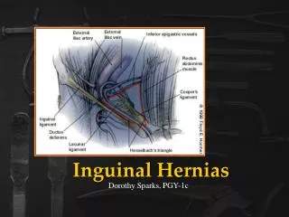
Inguinal hernia Inguinal Canal Anatomy • Anterior wall: • aponeurosis of external oblique (along entire length), • internal oblique on lateral one third • Posterior: • fascia transversalis • conjoint tendonon in medial one third • Roof: • arching fibers of internal oblique ,and • transversus abdominis • Floor (inferior): • inguinal ligament, and • lacunar ligamen at the medial end
Inguinal hernia Inguinal Canal Contents: • Male: • Spermatic cord structures: • vas deferens, • testicular artery • testicular veins (pampiniform plexus), • genital branch of genitofemoral nerve, • artery of the vas deference, • lymphatics, • autonomic nerves, • processus vaginalis. • Ilio inguinal nerve • Female: • Round ligament of the uterus, • genital branch of genitofemoral nerve, • lymphatics, • sympathetic plexus.
Inguinal hernia Signs & symptoms: • Bulge that enlarges when stand or strain, but often asymptomatic. • In general direct hernias produce fewer symptoms than indirect hernias and are less likely to complicate. • On examination: • Palpable defect or swelling may be present . • Indirect Hernia usually bulge at Internal Inguinal Ring. • Direct Hernia usually bulge at External Inguinal Ring.
Inguinal hernia There are two types of inguinal hernia: • Direct inguinal hernia • Indirect inguinal hernia
Differences between direct and indirect hernias • Origin and coarse: • Direct:Develops in the area of Hasselbach's triangle. The origin is medially to the inferior epigastric vessels. • Indirect: Develops at the internal ring. The origin is lateral to the inferior epigastric artery. • Content: • Direct:Retroperitoneal fat. less commonly, peritoneal sac containing bowel . • Indirect:Sac of peritoneum coming through internal ring, through which omentum or bowel canenter. • Etiology: • Direct: weakness of the posterior floor of the inguinal canal (acquired). • Indirect:patent processus vaginalis (Congenital) .
Differences between direct and indirect hernias • Boundaries of Hasselbach's triangle: • Medially: lateral border of rectus abdominis. • Laterally: inferior epigastric vessels. • Inferiorly: inguinal ligament.
Inguinal hernia • Differential diagnosis: • Tendonitis • Muscle tear • Lymphadenopathy • Lipoma • Varicose vein • Hydrocele • Epididymitis • Spermatocele
Inguinal hernia • Complications: • Irreducibility, but without signs of obstruction or strangulation • Small Bowel Obstruction, Usually urgent surgical repair • Strangulation, Surgical emergency 50% indirect, 3-10% direct.
Inguinal hernia Management: • Inguinal hernias should always be repaired ( herniotomy, herniorrhaphy ) unless there are specific contraindications. • Types of operations: • a permanent sutures, as in Shouldice repair (layered suture). • a permanent mesh -greater frequency to decrease tension.
Inguinal hernia management • Treatment of aggravating factors (chronic cough, prostatic obstruction, etc). • Use of truss (appliance to prevent hernia from protruding) when a patient refuses operative repair or when there are absolute contraindications to operation
Inguinal hernia • Both types (direct and indirect inguinal hernia) may occur at the same time and straddle the inferior epigastric artery. • This is called: Pantaloon hernia
Femoral hernia • The defect is in the transversalis fascia overlying the femoral ring at the entry to the femoral canal. • The hernia passes through the femoral canal and presents in the groin, below and lateral to the pubic tubercle. • It is more common in females and carries a higher risk of strangulation. • Femoral canal-ant.by inguinal ligament,post by fascia over pectineus muscle,lat. by femoral vein n medial by lacunar ligament
Femoral hernia Signs & symptoms: • A lump occurs below and lateral to the pubic tubercle. It may be reducible. • It may not be noticed until it becomes tender and painful. • This type of hernia should be carefully sought in the obese patient who presents with signs of intestinal obstruction without an obvious cause. • DD’s-saphena varix,enlarged inguinal LN,femoral artery aneurysm,rare femoral abscess.
Femoral hernia Surgical repair: • An incision is made directly over the swelling. • The sac is opened and the contents reduced and the sac removed. • Femoral canal obliterated with 3 interrupted non absorbable suture. • Treatment of strangulation or obstruction, if present. • There is no place for a truss in the treatment of femoral hernia.
Umbilical hernia • This occurs in children becauseof incomplete closure of the umbilical orifice. • The majority close spontaneously during the first year of life. • Surgical repair should only be carried out if the hernia has not disappeared by the age of 3 and the fascial defect is greater than 1.5cm in diameter.
Para-Umbilical hernia • It occurs just above or just below the umbilicus, and is more common in obese females. • Predisposing factors • multiple pregnancies and • obesity.
Para-Umbilical hernia • The neck of the sac is usually narrow and therefore there is a high risk of strangulation. • The most common content is • omentum ,then • transverse colon and small intestine. • Treatment: is by • Contents of sac freed from it’s wall,excision of the sac, and fascial defect repaired by • Upper flap overlapping the lower,a two layer overlapping repair thereby doubling the strength of repair (Mayo repair) • >4 cm,recurrent-polypropylene mesh
Epigastric hernia • This is usually a small protrusionthrough the linea Alba in the upper part of the abdomen. • It consists of : • extraperitoneal fat only, but • May contain omentum or small bowel.
Epigastric hernia • It may be extremely painful, probably because of trapping and ischaemia of extraperitoneal fat. • Treatment • is by enlaging the defect,excising the fat, simple suture of the defect with non-absorbable sutures . • >4 cm propylene mesh placed retromuscular plane
Incisional hernia • This occurs through a defect in the scar of a previous abdominal incision.
Incisional hernia • Etiology : • Age: Wound healing is poor in the older patient. • Obesity. • Postoperative wound infection. • Postoperative wound haematoma. • Raised intra-abdominal pressure postoperatively, e.g. coughing, straining, constipation, ileus. • Steroid therapy. • Type of incision: Midline vertical wounds have a higher incidence than transverse incisions. • Poor suturing technique: Rarely does a suture break
Incisional hernia • Sign & symptoms : • A swelling protrudes through the wound. • It May occur up to 5 years postoperatively. • Many are large and involve the whole incision and consequently the neck of the sac is wide and the risk of strangulation rare. • If the defect is small there is a greater risk of strangulation . • Treatment-palliative-abd.belt • - preoperative measures-reduce weight,treat cough,improve nutritional status.stop smoking. • -surgery:excision of sac,identification n apposition, • -large hernia-poly propylene mesh,
Richter’s hernia • Part of the wall of the intestine becomes trapped in the defect. • This is usually the antimesenteric border of the small bowel. • The lumen is intact ( no obstruction )
Diaphragmatic hernia • Traumatic: rare and followed by injuries to chest and abdomen. The Lt diaphragm is affected more than Rt and is accompanied by herniation of stomach and spleen. • Hiatus: • Sliding. • Para-esophegial.
Diaphragmatic hernia • Sliding: • in which the gastroesophogeal junction itself slides through the defect into the chest.
Diaphragmatic hernia • Para-esophageal • in which the junction remains fixed while another portion of the stomach moves up through the defect. • This can be dangerous as they may allow the stomach to rotate and obstruct.
Some other hernias • Spigelian hernia: • This is ahernia through the linea semilunaris at the lateral border of the rectus sheath. • Littre's hernia: • A hernia that contains a Meckel's diverticulum in the sac. • Obturator hernia: • This hernia occurs through the obturator foramen. It is commoner in elderly females. • Lumbar herniae: • These occur in the lumbar region (below the 12th rib & above the iliac crest).