Download
1 / 32
370 likes | 726 Vues
Prokaryotic microbial diversity. Taxonomy Study of the classification of living things Bacterial taxonomy has been difficult Species concept does not work Ideally, classify by evolutionary relationship Until recently, classification methods similar to identification methods

E N D
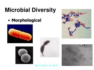
Prokaryotic microbial diversity • Taxonomy • Study of the classification of living things • Bacterial taxonomy has been difficult • Species concept does not work • Ideally, classify by evolutionary relationship • Until recently, classification methods similar to identification methods • Morphology, etc.: shape, size, arrangement • Gram stain • Biochemical characteristics, e.g. aerobe
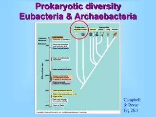
Relatedness is determined from DNA • Since 1960s, molecular techniques used to help determine relatedness • %G + C • DNA-DNA hybridization • Sequence of rRNA genes • Whole genome sequencing • rRNA gene sequences revealed the 3 domains • Archaea, Eubacteria, Eukaryota • Further genetic analysis shows family relationships • Gram negative and Gram positive difference is real.
Archaea: Archaebacteria • Different from Eubacteria because • rRNA genes different • Not peptidoglycan, but similar substance in cell walls • Odd branched lipids; other differences too. • Don’t cause disease • Classified into 3 groups by rRNA analysis • Some grow in normal environments, some are extremophiles. Examples of extremophiles: • Hyperthermophiles and acid loving species • Halophiles grow in very high NaCl concentrations • Methanogens: strict anaerobes, produce methane
Representative Eubacteria • Phototrophs • Autotrophic, use sunlight energy • Green sulfur, Green non-sulfur, Purple Sulfur, Purple non-sulfur • Bacteria use only 1 photosystem, do not produce oxygen • The “sulfur bacteria” can use reduced sulfur compounds as H donors for making sugar from CO2 • Color relates to pigments • Cyanobacteria: blue green “algae” • Similar to chloroplasts, release oxygen gas
Gram positive bacteria • Clostridium: rod shaped, spore forming • Strict anaerobes, ferment, make toxins • Mycoplasma: pleomorphic • Has no cell wall, stains pink, sterols in membranes • Bacillus: rod shaped spore former • Common in soil, some cause disease • Listeria: short rod • Psychrotrophic, causes disease • Lactobacillus: small fermenting rods • Helpful normal microbiota
Gram positive bacteria-2 • Streptococcus and Enterococcus • Round, fermenting, in chains, can cause disease • Staphylococcus: round, in clusters • Faculatative anaerobe, some cause disease • Corynebacterium: short rods in odd arrangements • Some normal microbiota, one causes disease • Mycobacterium: poorly staining rods • Have mycolic acids; cause disease or environmental • Actinomycetes: family of filamentous bacteria • Includes Streptomyces, maker of antibiotics
Gram Negative Bacteria • Wide variety with many families • 5 groups of “Proteobacteria” w/ Greek letters • Includes common environmental species, some that can’t make ATP, aerobes, anaerobes, disease causers • Pseudomonads: aerobic with great range of different C sources that can be used. • Enteric bacteria: facultative anaerobes, many of which can cause disease • Chlamydia: obligate parasites that cause disease • Spirochaetes: internal flagella, cause disease • Bacteroides and Cytophaga: much different genera with similar rRNA sequences.
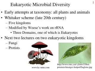
Eukaryotic Microbial Diversity • Early attempts at taxonomy: all plants and animals • Whitaker scheme (late 20th century) • Five kingdoms • Modified by Woese’s work on rRNA • Three Domains, one of which is Eukaryotes • Protista: the grab bag Kingdom • Always recognized as a highly diverse group • In new schemes, split into 7 kingdoms • Since the application of molecular biology, taxonomy of all things constantly changing.
Eukaryotes vs. prokaryotes • Eukaryotes are larger • Eukaryotes have membrane-bound organelles • Nucleus, mitochondria, membrane systems • Larger size requires functional compartments • Mitochondria once bacteria? So same size! http://www.earthlife.net/images/eury-cell.gif
Microbial eukaryotes • Animals • Parasitic worms; studied by Parasitologists • Fungi • Yeasts and molds, studied by Mycologists • Several types can cause human disease • Protists • Unicellular eukaryotes with many different characteristics. Also studied by Parasitologists. • Some cause human disease • Plants: not of particular interest other than hosts
Kingdom Protista • Highly diverse group of organisms • Size range from 5 µm to several meters (kelp) • Defined more by what they aren’t • Nutrient/energy acquisition ranges from photosynthesis to predatory to detrivores • Important in many food webs • Provide link between bacteria and larger organisms library.thinkquest.org/ 12413/protist.html
Some protozoal terminology • Macronucleus and micronucleus • Two type of nuclei differing in size and function. • Cyst: a resting stage similar to a spore with a thick wall and low level of metabolism. • Trophozoite: stage in life cycle during which the microbe is feeding and growing. • Merozoite: Small cells with a single nuclei produced during schizogony. • Large, multinucleated cell undergoes cytokinesis to produce multiple daughter cells (merozoites)
Plant-like Protists • Contain chloroplasts • Representatives • Diatoms (right). • Diatomaceous earth = fossilized diatoms: abrasives and slug repellants. • Red, brown, yellow algae • Seaweed, source of agar • Dinoflagellates • Neurotoxins and red tide http://www.bhikku.net/archives/03/img/diatoms.JPG www.enviroliteracy.org/ article.php/534.html
Fungus-like • Water molds • Slime molds Animal-like protists Capable of ingesting their food. Found among many different groups, so not good for taxonomy. http://en.wikipedia.org/wiki/Slime_mold http://ar.geocities.com/seti_argentina/estamos_solos/ameba.jpg
Breaking up the Protista: various algae, slime molds, and Protozoa • What are the characteristics of Protozoa? • Unicellular eukaryotes • Lack a cell wall • Require moist environments (water, damp soil, etc) • Great amounts of diversity • Locomotion: float, cilia, flagella, pseudopodia • Nutrition: chemoheterotrophs, photoautotrophs, either • Simple to complex life cycles, reproduction • Different cell organelles, some lack mitochondria
How to classify? • Cell ultrastructure and molecular analysis becoming the main methods used for classification. • Suggests that several kingdoms would be appropriate. • Alternative scheme, keep the kingdom Protista, classify protozoa into several phyla • Your text: • 4 groups of protozoa • Algae • Slime molds • Water molds
Classification of Protozoa Alveolates Ciliates Apicomplexans Dinoflagellates Amoebae Shelled and unshelled Euglenozoa Ameobae Euglenoids Kinetoplastids Archaezoa Diplomonadida Parabasala http://www.jracademy.com/~mlechner/archive1999/paramecium.JPG
Protozoa: details and examples • Alveolates • Possess alveoli: small membrane-bound cavities of unknown function (classification by ultrastructure) • Ciliates: move by cilia, short flagella-like appendages • Includes disease-causing Balantidium • Apicomplexans: have a complex of specialized organelles at the apices (corners, tips) of the cells • Generally have complex life cycles • Include Plasmodium (malaria), Toxoplasma (toxoplasmosis).
Apicomplexans Complex structure of organelles involved in infection. http://cgdc3.igmors.u-psud.fr/microbiologie/apicomplexans_fichiers/image002.jpg
Alveolates continued • Dinoflagellates • Large group of plantlike protozoa, have photosynthetic pigments (chlorophylls), cellulosic cell walls, store sugars as starch. • RNA sequences show relationship to other aveolates, not to plants. • Large portion of fresh water and marine plankton • Some encased in silica • Some bioluminescent or produce red pigments • Some produce dangerous neurotoxins
Amoebae • Amoebae have 2 main characteristics • Move and feed using pseudopodia • Cytoskeleton aids extension of cell membrane, cytoplasmic streaming. • Lack mitochondria • Some have loose shells; some form cysts. • Fossilized shells major component in some limestones. • Some “ameobae” are classified in another group. • Entamoeba: example of disease-causing amoeba.
Euglenozoa • United by similar RNA sequences • Not particularly similar otherwise. Have mitochondria. • Amoebae: move by pseudopodia • Including disease-causing Naegleria and Acanthameoba • Euglenoids: Euglena and similar microbes • Photoautrophs, but: no cell walls, motile by flagella and other means, store paramylon instead of starch, and can grow heterotrophically in the dark. Not plants! • Kinetoplastids: mitochondrial DNA forms kinetoplast • Includes Trypanosma, a pathogen
Archaezoa • Lack mitochondria and some other organelles • Thought to be old, hence the name (“Archae-”) • But have mitochondrial genes in nucleus. • Diplomonadida: 2 nuclei plus flagella • Includes pathogen Giardia, forms cysts, causes diarrhea • Parabasala: Single nucleus plus parabasal body. • Wood digesting microbe of termite gut. • Trichomonas, inhabits vagina, potential STD http://www.mhhe.com/socscience/sex/common/ibank/ibank/0149.jpg
Algae • Green algae • Ancestors of plants • Red algae • Mostly marine • Source of food thickeners carrageenan and agar • Chrysophyta (golden algae, diatoms, etc) • Diatoms: major component of phytoplankton • Diatomaceous earth as abrasives, gardening tools • Brown algae • Common seaweeds, kelps http://habitatnews.nus.edu.sg/news/chekjawa/ria/photos/r119.jpg
Water Molds and Slime Molds • Water molds • Similar to fungi except for 4 major differences; • 2 of 4: cellulose, not chitin in cell wall; motile spores • Phytophthora: Irish potato blight, sudden oak death • Slime molds • Acellular slime molds: The Blob, giant multi-nucleated cell; reproduces into amoebae that are amphibious • Cellular slime molds, e.g. Dictyostelium: unicellular, aggregate into slug-like structure, model for primitive development and differentiation.
Fungi • Mycology: the study of fungi • Fungi are mostly saprophytes, all heterotrophs • Saprophytes: decay non-living organic matter • Fungi are the kings of decomposition • Heterotrophs: use pre-formed organic matter • Not autotrophs, not photosynthetic • Fungi grow into, through their food • Release extracellular enzymes, break down polymers into LMW compounds for transport
Fungi terminology and structure • Hypha (singular) hyphae (plural): thread • Hyphae may be partially separated into cells or not at all (ceonocytic). • Cytoplasm is continuous throughout hypha • Mycelium (plural mycelia): a mass of hyphae • Like a bacterial colony except really all one organism. • Some fungi are molds, some are yeasts • Yeasts are oval, unicellular • Dimorphic: able to grow as either form. • Typical of some disease-causing fungi
Impacts of Fungi • Disease: mycosis (plural mycoses) • Superficial (on hairs, nails) • Cutaneous (dermatophytes, in skin (athlete’s foot) • Subcutaneous (deeper into skin) • Systemic (in deeper tissues, usually via lungs) • Opportunists: serious disease when immune system is depressed. • Antibiotic production • Penicillium, Cephalosporium • Decomposition; Food industry (soy sauce)
Classification of fungi • By sexual reproductive structures • Fungi reproduce both asexually and sexually • Deuteromycota = Fungi Imperfecti • No longer a valid classification • Contained fungi that couldn’t be coaxed into having sex • Through morphological and molecular means (e.g. DNA analysis), being distributed into the other 3 phyla of fungi.
Classification-2 • Zygomycota: produce zygospores • Example: Rhizopus • Fusion of hyphae (haploid) of opposite mating types produces zygospore (diploid). • Zygospore produces a zygosporangium with haploid spores that are released. • Asexually, sporangium containing spores. sporangia Zygospore botit.botany.wisc.edu/ images/332/Zygomycota/Z...www.butte.cc.ca.us/.../ fungi.unks.html
Classification-3 • Ascomycota: the sac fungi • Sexual spores produced inside an ascus (sac) • Asexual spores are called conidiospores or conidia (singular conidium) • Many types of common molds are ascomycetes. Ascus conidia fungus.org.uk/ nwfg/ascus.htm inseto.rc.unesp.br/.../ fungos%20e%20micoses.htm www.ent.iastate.edu/.../ aspergillus_ear_rot.html
Classification-4 • Basidiomycota: the club fungi or mushrooms • After extensive growth of hyphae, opposite mating types fuse and above ground mushroom is formed. • Sexual spores are called basidiospores; asexual conidia can also be formed. Close-up of gills www.birdsasart.com/ bn106.htm www.fishing-in-wales.com/. ../fungi/parasol.htm