Understanding Plasma Membrane Structure and Function: A Comprehensive Overview
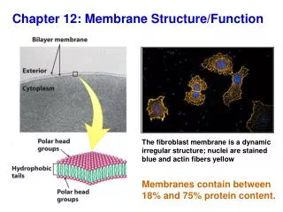
The plasma membrane is a selectively permeable barrier composed mainly of phospholipids and proteins. This membrane is flexible, allowing it to bend and change shape while supporting various macromolecules. The Fluid Mosaic Model explains its dynamic nature, where lipids and proteins move laterally, contributing to various cell functions like communication and transport. Integral and peripheral proteins assist in transporting substances and facilitating cell recognition. Cholesterol plays a crucial role in maintaining membrane fluidity, making it vital for cellular activities.

Understanding Plasma Membrane Structure and Function: A Comprehensive Overview
E N D
Presentation Transcript
Plasma Membrane • Plasma membrane is selectively permeable, (allowing some substances to cross more easily than others) • PM is flexible – bends and changes shape
Macromolecules in PM • Mostly lipids and proteins; some carbohydrates • Lipids are in the form of phospholipids (mostly) • Phospholipids are amphipathic molecules. • have both hydrophobic regions and hydrophilic regions.
hydrophilic extracellular Phospholipids … polar … are amphipathic nonpolar amphi- “on both sides” hydrophobic nonpolar polar intracellular hydrophilic
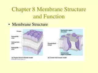
Membrane models- history • 1895 – PM is made of lipids (water interaction??) • 1925 – Phospholipid bilayer (flexibility; adhesion??) • 1935 – Davson & Danielli – proteins are outside phospholipid bilayer (hydrophobic proteins, variability???) 1950 - EM
Membrane models • Freeze-fracture studies (SEM) – proteins in the middle of PM • Fluid Mosaic Model – Singer and Nicholson (1972)
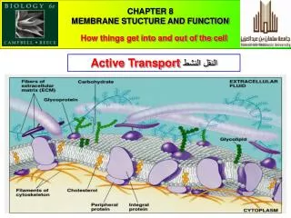
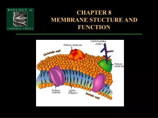
Fluid Mosaic model • Phopholipid makes a bilayer; Integral and peripheral proteins are found ‘within’ or on ‘one side’ of the phospholipid bilayer as the name indicates • Hydrophilic regions of proteins (polar side chains) and phospholipids (polar head)– in contact with water • Hydrophobic tails/nonpolar protein regions are in a nonaqueous environment - tucked away in the middle of the membrane • Weak hydrophobic interactions maintain membrane structure; cholesterol - maintains fluidity; • Glycolipids (antigens) found on outside side of the membrane - provide cell-cell recognition (immune function) • Ion channels are formed by integral proteins to transport polar substances • Some integral/peripheral proteins are enzymes or receptors or junctions - function in cell communication
Fluid Mosaic model Fig. 8.2b
Why ‘Fluid’ and Why ‘Mosaic’? Cholesterol (restricts it; except at low temp) Fluid Mosaic Proteins and lipids move Laterally and can also Flip Function is related to structures (AP Theme!); therefore function is a mosaic - elaborate… Structure is not the same on the inside and outside and from region to region (relate to lipids, integral and peripheral proteins, carbohydrates)
Membranes are fluid • Most of the lipids and some proteins can drift laterally in the plane of the membrane, but rarely flip-flop from one layer to the other. • Fluidity depends on unsaturation of fatty acids and presence of cholesterol. More unsaturation, more fluidity (why?). • At normal and high temp. cholesterol limits fluidity; at low temp. cholesterol actually prevents freezing of membrane (increases fluidity)
Membranes are mosaics of structure and function -Glycolipid (antigens) only on Extracellular side • A membrane is a collage of differentproteinsembedded in the fluid matrix of the lipid bilayer. • Peripheral proteins are loosely bonded to the surface (inside or outside) of the PM, often connected to the other membrane proteins. • Integral proteins penetrate the hydrophobic core of the lipid bilayer, often completely spanning the membrane (a transmembrane protein).
Proteins in Membranes Integral Proteins form alpha helix coils in the membrane Proteins are connected to cytoskeleton (intracellular) and Extra Cellular Matrix (outside)
Integral Transmembrane Protein: Asymmetry in amino acids gives the two membrane faces (extracellular and intracellular) their different characteristics
Mosaic Structure Cont’d • Membranes have distinctive inside and outside faces. • The two layers may differ in lipid composition, and proteins in the membrane have a clear direction. • The outer surface also has carbohydrates.
Mosaic Function • The proteins in the plasma membrane may provide a variety of major cell functions. Fig. 8.9
Provides a hydrophilic channel that is selective for a particular solute Campbell; Fig 8.9
“active site” “conformational change”
Membrane carbohydrates are important for cell-cell recognition • important in tissue and organ development. • rejection of foreign cells by the immune system. • Carbohydrates are covalently bonded either to lipids, forming glycolipids, or, more commonly, to proteins, forming glycoproteins • Example: The four human blood groups (A, B, AB, and O) differ in the external carbohydrates on red blood cells
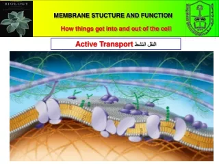
Transport Across the Membrane • Small molecules and ions moves across the plasma membrane in both directions. Wastes, ions, CO2 Sugars, amino acids, oxygen, ions
Hydrophobic molecules (lipids): hydrocarbons, CO2, and O2 Small polar molecules - water Can get through because of small size or hydrophobic nature Ions and polar molecules like glucose, amino acids Size + charged nature prevents easy acess When do these substances need to get in? Easy Access Need Assistance
Proteins can assist andregulate the transport of ions and polar molecules
Energy Requirements Requires no energy other than that of molecular motion Passive Transport Diffusion Facilitated Simple Molecules go through lipid bilayer Requires TRANSPORT proteins Channel Proteins Carrier Proteins
Passive transport- 1) Simple Diffusion • Diffusion is the tendency of molecules of any substance to spread out in the available space • driven by the intrinsic kinetic energy of molecules (so temp increase will…….). • a substance will diffuse from where it is more concentrated to where it is less concentrated, i.e. down its concentration gradient. O2, and CO2; small polar molecules include ethanol, H2O, and urea.
Passive transport- 2) Facilitated Diffusion • 1) Channelproteins -provide corridors allowing a specific molecule or ion to cross the membrane. • These channel proteins allow fast transport • Water channel proteins -aquaprorins • Ions move through these channels
(SKIP details) Channels Ions, and H2O Open Closed Channels that spend almost all of their time in the open configuration are called “leak” channels, or pores Channels that spend almost all of their time in the closed configuration are called “gated”
Passive transport- 2) Facilitated Diffusion 2) Carrier Proteins – Allows movement of polar compounds • Molecules (substrates) actually bind with the carrier (amino acids, sugars, nucleosides, and other small molecules). Know this - ‘glucose’ and ‘aminoacid’ carrier proteins transport these substances into the blood from the small intestine! • 2. Protein changes in shape (conformational change) • 3. It allows molecules to pass through
Transport Proteins are like Enzymes • Transport proteins Enzymes • Specific binding sites • Can become saturated • Can be inhibited • Catalyze a process
3.Osmosis is the passive transport of water Tonicity describes how the size of a cell would change if it were placed in a solution
Osmosis is the passive transport of water – it occurs until isotonicity is reached Osmosis – passive diffusion of water across a semipermeable membrane Fig. 8.11
Osmosis is the diffusion of water through a selectively permeable membrane. Tonicity describes how the size of a cell would change if it were placed in the solution Isotonic – same solute/water concentrations as inside cells so cells retain their normal size and shape
hypertonic H2O hypotonic H2O Lab - hemodialysis animation
Be sure to know how these calculations were performed (on next test). Open system; osmosis raises water in arm with more solute Plunger pushes and opposes osmosis (back pressure) Plunger keeps pushing, causing water to move against concentration gradient Result: Water rises in arm on opposite side
Wilted (cells may be flaccid or plasmolysed) Turgid
Cell survival depends on balancing water uptake and loss • Osmoregulation : maintanance of osmotic balance • Paramecium – protist (contractile vacuole)
The cells of plants, prokaryotes, fungi, and some protists have cell walls that contribute to the cell’s water balance.
4.Active transport is the pumping of solutes against their gradients • Active transport requires the cell to use its own metabolic energy (ATP). • Active transport is against the Concentration Gradient (solutes move from low concentration to high concentration)
The sodium-potassium pump actively maintains the gradient of sodium (Na+) and potassium ions (K+) across the membrane.
Some ion pumps generate voltage across membranes • All cells maintain a voltage across their plasma membranes using pumps. • The cytoplasm of a cell is negative in charge compared to the extracellular fluid because of an unequal distribution of cations (+) and anions (-) on opposite sides of the membrane. • This voltage, the membrane potential, ranges f50 to -200 millivolts.
The membrane potential acts like a battery. • The membrane potential favors the passive transport of cations (+) into the cell and anions (-) out of the cell. • Two combined forces, collectively called the electrochemical gradient, drive the diffusion of ions across a membrane: • a chemical force based on an ion’s concentration gradient • an electrical force based on the effect of the membrane potential on the ion’s movement.
Some ion pumps generate voltage across membranes • Ions diffuse down their electrochemical gradient. • Special transport proteins, electrogenic pumps, generate the voltage gradients across a membrane • Examples of electrogenic pumps – Na-K pump, proton (H+) pump
Proton pump actively transports H+ out of the cell. • Plants, fungi, mitochondria, chloroplast • These electrogenic pumps store energy that can be used to make ATP.
In cotransport, a membrane protein couples the transport of two solutes oncentration gradient.
Exocytosis and Endocytosis transport large molecules (need ATP) • Small molecules and water Transport proteins • Large macromolecules Vesicles • Exocytosis: a transport vesicle budded from the Golgi apparatus is moved by the cytoskeleton to the plasma membrane.