Download
1 / 26
570 likes | 1.59k Vues
Classification of vascular anomalies. Updated ISSVA (International Society for the Study of Vascular Anomalies). Department of diangosis imaging Children’s Hospital 2. INTRODUCTION. Vascular anomalies are among the most common congenital abnormalities observed in infants and children.

E N D
Classification of vascular anomalies Updated ISSVA (International Society for the Study of Vascular Anomalies) Department of diangosis imaging Children’s Hospital 2
INTRODUCTION • Vascular anomalies are among the most common congenital abnormalities observed in infants and children. • Older nomenclature continues to cause confusion, misunderstood diagnoses, and potential mismanagement • In 1982, Mulliken and Glowacki proposed a classification system for vascular anomalies based on their clinical behavior and endothelial cell characteristics into two groups: hemangiomas and vascular malformations.This system, which was adopted by the ISSVA, has since been expanded and is now widely accepted. • Radiologists can use the ISSVA classification system by correlating imaging findings with patient history and physical findings. Consistent use of this system will help patients receive the correct diagnosis and treatment.
Traditional classification • Capillary hemangioma • Strawberry hemangioma • Strawberry nevus • Port wine stain • Flame nevus • Cavernous hemangioma • Venous angioma • Lymphangioma • Arteriovenous malformation
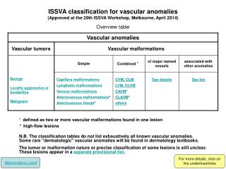
Updated ISSVA classification of vascular anomalies. Vascular tumors Vascular malformations • Infantile hemangiomas • Congenital hemangiomas (RICH and NICH) • Tufted angioma (with or without Kasabach-Merritt syndrome) • Kaposiform hemangioendothelioma (with or without Kasabach-Merritt syndrome) • Spindle cell hemangioendothelioma • Other, rare hemangioendotheliomas (epithelioid, composite, retiform, polymorphous, Dabska tumor, lymphangioendotheliomatosis, etc.) • Dermatologic acquired vascular tumors (pyogenic granuloma, targetoid hemangioma, glomeruloid hemangioma, microvenular hemangioma, etc.) 1..Slow-flow vascular malformations: • Capillary malformation (CM) • Port-wine stain • Telangiectasia • Angiokeratoma • Venous malformation (VM) • Common sporadic VM • Bean syndrome • Familial cutaneous and mucosal venous malformation (VMCM) • Glomuvenous malformation (GVM)(glomangioma) • Maffucci syndrome • Lymphatic malformation (LM) 2. Fast-flow vascular malformations: • Arterial malformation (AM) • Arteriovenous fistula (AVF) • Arteriovenous malformation (AVM) 3.Complex-combined vascular malformations: • CVM, CLM, LVM, CLVM, • AVM-LM, CM-AVM C:capillary; V:venous; L:lymphatic; AV:arteriovenous; M:malformation. RICH:rapidly involuting congenital hemangioma; NICH:noninvoluting congenital hemangioma.
Comparison of Previous Terminology and New ISSVA Terminology
Key Imaging Features of the Most Common Pediatric Vascular Anomalies
Differentiation between Hemangioma and Hemangioendothelioma of the liver
Classification of Vascular Tumors • Benign tumors and tumor-like conditions • Hemangiomas • Spindle cell hemangioma (‘hemangioendotheliomas’) • Epithelioid hemangioma • Low-grade malignant tumors • Retinform hemangioendotheliomas • Composite hemangioendotheliomas • Polymorphous hemangioendotheliomas • Kaposiform hemangioendotheliomas • Malignant tumors • Epithelioid hemangioendotheliomas • Angiosarcoma
HEMANGIOMA Arterio-Venous Malformation(AVM) congenital hemangioma Infantile hemangioma
HEMANGIOMA Kaposiform Hemangioendothelioma with Kasabach-Merritt Phenomenon
Hemangioma • Benign endothelial cell tumor • 2 main types 1. Infantile Hemangioma • Most common tumor of infancy/childhood • Usually has overlying patch of redness • Appears weeks/months after birth • Natural course - 3 stages 1. Proliferating - first year 2. Involuting - few years 3. Involuted - most resolved by age 10
Hemangioma(cont) 2. Congenital Hemangioma • Present at birth • Rare (compared to infantile) • Blue/gray hue w/ pale halo (skin) • 2 types • Non-Involuting (NICH) - persistent • Rapidly Involuting (RICH) - resolved by 1-2 yrs
Lymphatic malformations • Commonly occur in the cervicofacial region, Lymphatic malformations in an extremity can cause diffuse or localized swelling with soft-tissue and skeletal overgrowth
Venous Malformation (VM) • Present at birth • skin discoloration, local swelling, and pain • Thin-walled, dilated veins:Inadequate smooth muscle layer • Complications: Thrombosis, bleeding
Arterio-Venous Malformation (AVM) • Present at birth • Reddish vascular hue (skin), often warm pulsations, thrill, and bruit • High-flow arterio-venous communication - absence of developed capillary bed • Complications: ulceration, bleeding,pain, compression/displacement of organs, high-outputcardiac failure
Summary of Regional and Diffuse Syndromes Associated With Vascular Malformations • Regional syndromes with associated vascular malformations • Sturge–Weber: facial capillary malformation with intracranial capillary malformation, venous malformation, or AVM. • Klippel–Trenaunay: limb/trunk capillary venous lymphatic malformations with overgrowth. • Parkes Weber: CAVM with overgrowth; lymphatic malformation. • Diffuse syndromes associated slow-flow malformations • Proteus syndrome: vascular malformations (capillary or venous), hamartomatous syndrome with overgrowth(hemihypertrophy and macrodactyly), lipomas, pigmented nevi. • Blue rubber bleb nevus (Bean) syndrome: multiple cutaneous, musculoskeletal, and gastrointestinal tract venous malformations. • Epidermal nevus syndrome (Solomon syndrome): vascular malformations (intracranial AVM), epidermal nevi, various developmental abnormalities of the skin, eyes, nervous, skeletal, cardiovascular, and urogenital systems. • Bannayan–Riley–Ruvalcaba syndrome: vascular malformations (cutaneous, intracranial), macrocephaly, ectodermal dysplasia, lipomatous masses, and intestinal hamartomatous polyps, PTEN suppressor gene mutation association. • Diffuse syndromes associated fast flow malformations • Hereditary hemorrhagic telangiectasia (Osler–Weber–Rendu): telangiectasias (skin, mucous membranes, gastrointestinal mucosa) and AVMs (lungs, liver, brain, spinal cord). AVM, arteriovenous malformation; CAVM, capillary arterial venous malformation; PTEN, phosphatase and tensin homolog.
OVERGROWTH SYNDROMES: • Klippel-Trénaunay syndrome which is a low-flow combined vascular anomaly (capillary-lymphatic-venous malformation) usually associated with marked overgrowth of the leg and capillary stains. • Parkes-Weber syndrome consists of an AVM-like high-flow malformation that involves the entire extremity (usually a lower limb), and it is usually associated with a capillary malformation over the enlarged limb.
Simple malformations • slow flow • capillary • lymphatic • venous • fast flow • arterial • aneurysm • coarctation • ectasia • arteriovenous fistulae (with one ore more shunts) • arteriovenous malformations (with a nidus of multiple shunts)
Complex malformations • regional • Sturge-Weber syndrome • Klippel-Trénaunay syndrome • F. P. Weber syndrome • diffuse • Maffucci syndrome • Solomon syndrome • Proteus syndrome
Low flow malformations • Lymphatic Malformations -Microcystic -Macrocystic • Venous Malformations • Capillary Malformations • Combined types