Download
1 / 79
790 likes | 1.12k Vues
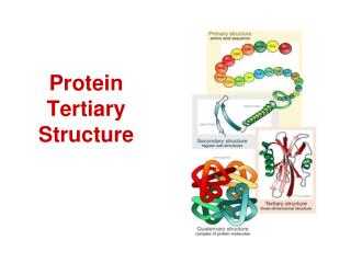
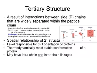
Domain,tertiary , and quarternary structure of proteins. Levels of protein structure organization. Between secondary and tertiary structure. Supersecondary structure : arrangement of elements of same or different secondary structure into motifs ; a motif is usually not stable by itself.

E N D
Between secondary and tertiary structure • Supersecondary structure: arrangement of elements of same or different secondary structure into motifs; a motif is usually not stable by itself. • Domains: A domain is an independent unit, usually stable by itself; it can comprise the whole protein or a part of the protein.
Example of a protein with two b-hairpins: erabutoxin from whale. Example of a b-hairpin in bovine pancreatic trypsin inhibitor– BPTI.
Helix E helix F Troponin C with four EF motifs that bind calcium ions. Because of high content of acidic amino-acid residues with side chains pointing inside the loop, the EF-hand motif constitutes a calcium-binding scaffold in troponin, calmodulin, etc.
The Helix-Turn-Helix motif • This motif is characteristic of proteins binding to the major DNA grove. • The proteins containing this motif recongize palindromic DNA sequences. • The second helix is responsible for nucleotide sequence recognition.
b-a-b Motif (very important and very frequent) Hydrophobiccorebetweena-helix and b-sheet
Domains: classification criteria • Functionality (performing a biological function or role in formation and stabilization of globular structure) • Solubility: • Globular proteins and protien domains (water solubke) • Membrane proteins and domains (lipid soluble) • Fibrillar protiens (insoluble) • Content of secondary structure • aa (parallel and antiparallel) • bb • a/b • a+b • high disulfide-bridge or metal content.
Protein domains Sposób wyróżnienia domeny w cząsteczce białka jest często intuicyjny, ale możliwe jest przypisanie domenom pewnych, wyróżniających je cech: - domena jest potencjalnie niezależną jednostką fałdowania, - domena jest lokalną, zwartą, globularną, półniezaeżną częścią białka, związaną z nim jednak kowalencyjnie, - sekwencją aminokwasów, charakterystyczną dla danej domeny, można spotkać w innych, podobnych domenach tego samego białka (lub w innych białkach), - domenom towarzyszą często specyficzne funkcje (np. wiązanie nukleotydów, sacharydów) - przestrzeń między domenami wyznacza często centrum aktywne białka, - domena reprezentuje zwarty, genetyczny segment (np. domeny w immunoglobulinach, dehydrogenazach, globinach) Pojedyńcza cząsteczka białka może posiadać kilka lub więcej domen ale większość białek należy do grupy białek jednodomenowych.
Domains: example • Domains of recently evolved proteins are frequently encoded by exons, reflecting gene fusion of simpler modules. For example, in the case of hepatocyte growth factors and plasminogens, a number of kringle domains are present. • For a recent review on domain insertion. Domain swapping between two protomers is not uncommon (for example in the case of diphtheria toxin). • Domains form an important level in the hierarchical organisation of the three-dimensional structure of globular proteins, although not all proteins can be described as multidomain structures.
Example if division of a protein into domains: human Hsp70 chaperone
Domain identification algorithms Schultz’s method: neighborhood correlation criterion
The Go algorithm: interdomain distances are larger than intradomain distances
The Rose algorithm: based on the deviation of the long axes of the fragments from protein mean plane; works for continuous domains
The Crippen algorithm: based on dissection of residues according to interresidue distances into clusters Ca-Ca distances between secondary structures are represented in the form of average values termed 'proximity indices' and the secondary structural organisation is indicated in the form of dendrograms. An example is shown for the case of calmodulin.
Specific nodes in these dendrograms are identified as tertiary structural clusters of the protein; these include supersecondary structures and domains. A ratio of the average proximity indices (ignoring inter-clusteral distances) to the average of all proximity indices, weighted for the aggregation of small sub-clusters and termed the disjoint factor, is employed as a discriminatory parameter to identify automatically clusters representing individual domains. An example of domains identified in glutathione reducatase is shown below :
The domains identified by this clustering method may not correspond to the functional domains proposed. The "disjoint factor" gives a measure of the extent of interaction between domains and has been used to classify domains into one of the three types, disjoint, interacting and conjoint. Domains are classified as those with sparse inter-domain interfaces (disjoint), intermediate interactions (interacting) and elaborate interfaces (conjoint) based on the magnitude of the disjoint factor. An example of the three types is shown below :
Classification of three-dimensional structures of protein Richardson’s classification a– a-helices are only or dominant secondary-structure elements (e.g., ferritin, myoglobin) b – b-sheets are only or dominant elements (e.g., lipocain) a/b – contain strongly interacting helices and sheets a+b – contain weakly interacting or separated helices and sheets
SCOP classification • Structural Classification Of Proteins • This is a hierarchical classification scheme with the following 4 levels: • Families – one family is comprised by proteins related structurally, evolutionally, and functionally. • Superfamoilies – A superfamily is comprised by families of substantially related by structure and function. • Folds – Superfamilies with common topology of the main portion of the chain. • Classes - Groups of folds characterized by secondary structure: a (mainly a-helices), b (mainly b-sheets), a/b (a-helices and b-sheets strongly interacting), a+b (a-helices and b-weakly interacting or not interacting), multidomain proteins (non-homologous proteins with vert diverse folds).
Scop Classification Statistics SCOP: Structural Classification of Proteins. 1.75 release38221 PDB Entries (23 Feb 2009). 110800 Domains. 1 Literature Reference(excluding nucleic acids and theoretical models)
CATH classification (Class (C), Architecture(A), Topology(T), Homologous superfamily (H)) • Four hierarchy levels: • Class (Level C): according to the content of secondary structure type a, b, a&b (a/b and a+b), weakly or undefined secondary structure. • Architecture. (Level A) – Orientation and connection topology between secondary structure elements. • Topology. (Level T) – based on fold type. • Homoloous superfamilies. (Level H) – high homology indicating a common anscestor: • >30% sequence identity OR • > 20% sequence identiy and 60% structural homology OR • > 60% structural homology and similar domains have similar function.
Class(C)derived from secondary structure content is assigned automatically • Architecture(A)describes the gross orientation of secondary structures, independent of connectivity. • Topology(T) clusters structures according to their topological connections and numbers of secondary structures • Homologous superfamily (H) [ http://www.biochem.ucl.ac.uk/bsm/cath_new/ ]
A „periodic table” of protein structures W. Taylor, Nature, 416, 6881, 657-660 (2002)
a-helical structures ROP: two packed helices 1rop - RASMOL
Antiparallel four-a-helix bundle 15o twist of helix axes
Example: ferritin 1fha - RASMOL