Basic Structure of Proteins
430 likes | 691 Vues
Understand the primary, secondary, tertiary, and quaternary structures of proteins and their biological significance. Explore how proteins function in essential biological processes like enzyme catalysis, molecular transport, movement, sensory reception, immunity, and cellular functions. Learn how the amino acid sequence determines protein structure.

Basic Structure of Proteins
E N D
Presentation Transcript
Basic Structure of Proteins Primary, Secondary, Tertiary & Quaternary Structure of Proteins
THINGS YOU MUST KNOW • How many structures does a protein have? • What is the primary structure of protein? • Which is the biological importance of the primary structure of proteins? • Which is the stabilizing chemical bond of primary structure of proteins? • What is the secondary structure of proteins? • Which is the stabilizing chemical bond of secondary structure of proteins? • List the different types of models of secondary structure of proteins • Describe the a-helix model • Describe the b-pleated sheet model • Describe the b-turn model • List the analogies and differences between a-helix and b-pleated sheet structures • What is the tertiary structure of protein? • Which are the stabilizing chemical bonds of tertiary structure of proteins? • What is the importance of the tertiary level on protein structural organization? • What is the quaternary structure of proteins? • Which are the stabilizing chemical bonds of quaternary structure of proteins? • What is the importance of quaternary structure of proteins?
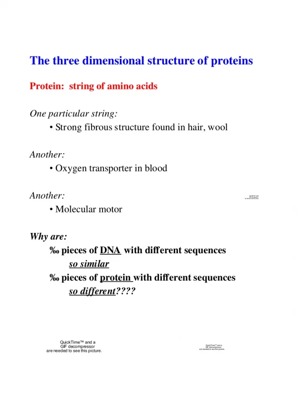
Proteins play a key role in biological processes. They can fulfill a vast variety of tasks. • Enzymes catalyze the complex set of chemical reactions that are collectively referred to as life. These chemical reactions are on the other hand regulated by proteins, which act either directly as components of enzymes or indirectly in the form of chemical messengers, or their receptors. • Proteins are engaged in the transport and storage of biologically important substances such as metal ions, oxygene, glucose, lipids, and many other molecules. • In the form of muscle fibers and other contractile assemblies, proteins generate the coordinated mechanical motion of numerous biological processes, including the separation of chromosomes during cell division and the movement of your eyes as you read this text. • Proteins such as rhodopsin in the retina of your eye, acquire sensory information that is processed through the action of nerve cell proteins. • The proteins of the immune system, such as the immunoglobulins, form an essential biological defense system in higher animals. • Proteins carry out a wide range of functions in the cell. This variety in capability is the result of the range of structures found in proteins and the wide variety of molecules proteins can bind with high specificity. • All of this results from the peculiar structure of proteins
Protein function can be understood only in terms of proteins structure. That means the structure of a protein determines its biochemical function. • Amino Acid Sequence Determines Primary Structure • When the number, structure, and order of all of the amino acid residues in a polypeptide are known, its primary structure has been determined. • This is the subject of this lecture.
In order to identify and classify known and new protein structures it has been found useful to describe the structure of a protein in terms of four levels.
Primary structureisthelinear sequence of amino acids in the polypeptide chain(s) of a protein. • Secondary structure-the local spatial arrangement of a polypeptides backbone atoms without regard to the conformations of its side chains. Secondary Structure consists of local regions of poly peptide chains that have a regular conformation (- helices, - sheets etc) which is stabilized by H-bonds. • Tertiary structure - the overall arrangement of secondary structure elements. • Tertiary Structure refers to the 3-D configuration of an entire polypeptide chain .This includes - helices & - sheets and regions that are globular or spherical • Quaternary structure-the arrangement of several polypeptide chains • Quaternary Structure consists of number of polypeptide chains or subunits joined by noncovalent interactions.
4 organizational levels: primary, secondary, tertiary and quaternary
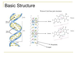
PRIMARY LEVEL OF PROTEIN STRUCTURAL ORGANIZATION It’s the sequence of amino acids linked through peptide bonds (Covalent backbone of the protein) H2N-Glu-Ala-Val-Ser-Leu-Ala-Lys-Cys-COOH H2N-Ala-Glu-Val-Ser-Ala-Leu-Lys-Cys-COOH
Primary Structure of Proteins The particular sequence of amino acids that is the backbone of a peptide chain or protein Ala-Leu-Cys-Met
The particular sequence of amino acids in a peptide or protein is referred to as the primary structure. For example, a hormone that stimulates the thyroid to release thyroxine consists of a tripeptide Glu-His-Pro.
Although other sequences are possible for these three amino acids, only the tripeptide with the Glu-His-Pro sequence of amino acids has hormonal activity. Sequences such as His-Pro-Glu or Pro-His-Glu do not produce hormonal activity. Thus the biological function of peptides as well as proteins depends on the order of the amino acids.When cells are damaged, a polypeptide called bradykinin is released at the site,which stimulates the release of prostaglandins. The presence of bradykinin, whichcontains nine amino acids, regulates blood pressure.
Primary structure - the amino acid sequence of the proteins polypeptide chains. • Proteins are linear polymers of 20 different amino acids linked by covalent amide bonds, called peptide bonds in a specific sequence of its constituent amino acids. • The sequence of amino acids that make up a protein is called its primary structure. • The Primary structure defines the linear sequence of amino acids and is due to the peptide bond . The order of the amino acids is called the amino acid sequence. The sequences of all polypeptide chains of a protein is defined as the primary structure. • The primary structure of a protein determines the three-dimensional structure and the function of the protein. • Peptide bonds are formed between the carbon atom (C) of the carboxyl group and the nitrogen atom (N) of the amino group of an amino acid. • Another way of saying this is that peptide bonds are formed by a condensation reaction between the amine group of one amino acid and the carboxyl group of another resulting in an amide group. The elements of water are removed as a by product of this reaction. Water (HOH) forms from the -OH of the carboxyl group of one amino acid and a hydrogen from the -NH2 group of the other amino acid. The product is called a peptide. Thus both peptides and proteins have amino and carboxyl ends.
The amino acids can be linked in any order, but the order of amino acids is unique to a given protein and is referred to as that protein's "primary structure." • The repeating sequence of “C-N-C-C-" resulting from amide bond formation is called the "polypeptide backbone." • Thedirectionality of the linkage results in directionality in the resulting polypeptide chain. The amino group on the first amino acid is referred to as the protein's "N-terminus" and the carboxyl group on the last amino acid is the "C-terminus." • Remember: • Primary structure COVALENT PEPTIDE BONDS • The protein backbone is represented as "C-N-C-C-". • Peptide bonds are formed between the carbon atom (C) of the carboxyl group and the nitrogen atom (N) of the amino group of an amino acid • The structure of a protein determines its biochemical function • The amino acids can be linked in any order but the order of amino acids is unique to a given protein and is referred to as that protein's "primary structure."
SECONDARY LEVEL OF PROTEIN STRUCTURAL ORGANIZATION It’s the spatial arrangement of amino acids inside the peptide chain stabilized through hydrogen bonds between the elements of the peptide bond. (Interactions of neighboring amino acids)
Secondary Protein Structure • Polypeptides form regular arrangements of amino acids called secondary structure • The secondary structure of a protein is the local spatial arrangement of a polypeptides backbone atoms without regard to the conformations of its side chains. • The secondary structure of a protein describes the way the amino acids next to or near to each other along the polypeptide are arranged in space. • In order for proteins to fold into their characteristic three-dimensional "conformations" the protein's "secondary structure" is adopted by hydrogen bonding between the N-H and C=O groups along the backbone or primary structure of the protein. • The three most common types of secondary structure are the alpha helix, the beta-pleated sheet (parallel or anti-parallel), and the triple helix found in collagen. • These patterns result from regular hydrogen bond patterns of backbone atoms in which the polypeptide chain spirals around a central "helix axis" with a clockwise twist. Thus the secondary structure of proteins refers to regular, repeated patters of folding of the protein backbone to create the distinctive structures shared by many proteins.
In each type of secondary structure, we will look at the hydrogen bonding between the hydrogen atom of an amino group in the polypeptide chain and the oxygen atom of the carboxyl group in another part of the chain.
α-Helix • Most common secondary structure. • Three-dimensional arrangement of amino acids with the polypeptide chain in a corkscrew shape or spiral . Looks like a coiled “telephone cord” • The a helix is a rodlike structure. The tightly coiled polypeptide main chain forms the inner part of the rod, and the side chains extend outward in a helical array off the long axis as shown in the slides that follow. • The a helix is held together and stabilized by hydrogen bonds between the NH and CO groups of the main chain. The CO group of each amino acid is hydrogen bonded to the NH group of the amino acid that is situated four residues ahead in the linear sequence in the next turn of the helix • To repeat H bonds are formed between the H of –N-H group of one amino acid and the –O of C=O of the fourth amino acid along the chain • Because many hydrogen bonds form along the peptide backbone, this portion of the protein takes the shape of a strong, tight coil that looks like a telephone cord. • All the side chains (R groups) of the amino acids are located on the outside of the helix. • α-Helix is found in • Keratins – almost completely helical; major component of hair, skin • · Rigidity determined by no. of –S-S- bonds • · Hb – 80% helical so globular, flexible molecule
In the alpha helix, all the main-chain CO and NH groups are hydrogen bonded. Each residue is related to the next one by a translation of 1.5 A along the helix axis and a rotation of 100 degrees, which gives 3.6 amino acid residues per turn of helix. • Thus, amino acids spaced three and four apart in the linear sequence are spatially quite close to one another in an a helix. • In contrast, amino acids two apart in the linear sequence are situated on opposite sides of the helix and so are unlikely to make contact. • The pitch of the a helix is 5.4 A, the product of the translation (1.5 A) and the number of residues per turn (3.6). • The screw-sense of a helix can be right-handed (clockwise) or left-handed (counterclockwise); the a helices found in proteins are right-handed. • The a-helix content of proteins of known three-dimensional structure is highly variable. In some, such as myoglobin and hemoglobin, the a helix is the major structural motif. • Other proteins, such as the digestive enzyme chymotrypsin, are virtually devoid of a helix. The single-stranded a helix is usually a rather short rod, typically less than 40 A in length. • A variation of the a-helical theme is used to construct much longer rods, extending to 1000 A or more. Two or more a helices can entwine around each other to form a cable. Such a-helical coiled coils are found in several proteins: keratin in hair, myosin and tropomyosin in muscle, epidermin in skin, and fibrin in blood clots. The helical cables in these proteins serve a mechanical role in forming stiff bundles of fibers.
Secondary structure includes various types of local conformations in which the atoms of the side chains are not involved. • An -helix is generated when each carbonyl of a peptide bond forms a hydrogen bond with the -NH of a peptide bond four amino acid residues further along the chain. • The side chains of the amino acid residues extend outward from the central axis of the rod-like structure. • The -helix is disrupted by proline residues, in which the ring imposed geometric constraints, and by regions in which numerous amino acid residues have charged groups or large, bulky side chains.
a-HELIX It’s the secondary level of protein organization in which the polypeptide backbone is tightly wound around an imaginary axis as an spiral structure. (Helicoidal arrangement of the peptide chain)
CHARACTERISTICS OF a-HELIX It is a rod-like structure Amino terminus The polypeptide chain is folded into a helix around a common axis There are about 3,6 amino acid residues per turn of the helix Each residue is linked to residues in the preceding and following turns by hydrogen bonds between the N-H groups and the oxygen atom of the C=O group Each amino acid establishes a hydrogen bond with other situated 4 residues ahead the helix The side chains R of the different residues project radially from the helix Carboxyl terminus
TYPES OF a-HELIX Left-handed Right -handed Right-handed a-helix is much more stable than left handed a-helix Essentially all a-helix found in proteins are right-handed
Secondary Structure – Beta Pleated Sheet • Another important periodic structural motif, or type of secondary structure is known as the beta-pleated sheet. • In a beta-pleated sheet, polypeptide chains are held together side by side by hydrogen bonds between the peptide chains. • For example, in a beta-pleated sheet of silk fibroin, the small R groups of the prevalent amino acids, glycine, alanine, and serine, extend above and below the sheet. • This results in a series of /3-pleated sheets that can be stacked close together. The hydrogen bonds holding the /3-pleated sheets tightly in place account for the strength and durability of fibrous proteins such as silk.
Comparison of β-sheet with α-helixThe beta pleated sheet differs markedly from the a helix in that it is a sheet rather than a rod. • The polypeptide chain in the beta pleated sheet is almost fully extended as shown in the slides to follow rather than being tightly coiled as in the a helix. • The axial distance between adjacent amino acids is 3.5 A, in contrast with 1.5 A for the a helix. • Another difference is that the beta pleated sheet is stabilized by hydrogen bonds between NH and CO groups in different polypeptide strands, whereas in the a helix the hydrogen bonds are between NH and CO groups in the same polypeptide chain. • Adjacent strands in a beta pleated sheet can run in the same direction (parallel beta sheet) or in opposite directions (antiparallel beta sheet). • H-bonds perpendicular to long axis • β-sheets are composed of two or more polypeptide chains or extended segments of thesame polypeptide
Secondary Structure – Beta Pleated Sheet • Polypeptide chains are arranged side by side • Hydrogen bonds form between chains • R groups of extend above and below the sheet • Typical of fibrous proteins such as silk
Secondary structure includes various types of local conformations in which the atoms of the side chains are not involved. • -Sheets are formed by hydrogen bonds between two extended polypeptide chains or between two regions of a single chain that folds back on itself. • These interactions are between the carbonyl of one peptide bond and the -NH of another. • The chains may run in the same direction or in opposite directions.
b-PLEATED SHEET • It’s the secondary level of protein organization in which the backbone of the peptide chain is extended into a zigzag arrangement resembling a series of pleats. • Disposition of the peptide bonds in planes of alternating slopes
CHARACTERISTICS OF b-PLEATED SHEET It is an extended structure made with different b-strands The planes of consecutive peptide bonds have different slopes (alternating ascending and descending direction) The side chain of adjacent amino acids point in opposite direction Hydrogen bonds link one b-strand with other and are perpendicular to the long axis of the sheet
TYPES OF b-PLEATED SHEET Antiparallel Parallel Antiparallel is more stable than parallel Both models are found in proteins
In some proteins, the polypeptide chain consists of mostly the a-helix secondary structure, whereas other proteins consist of mostly the /3-pleated sheet structure. Another group of proteins have a mixture with some sections of the polypeptide chain in a-helixes and other sections in the /3-pleated sheet structure. The tendency to form a certain type of secondary structure depends on the amino acids in a particular segment of the polypeptide chain. The amino acids that tend to form an a- helix or beta-pleated sheet follow.
Remember: • Secondary structure is HYDROGEN BONDS -- hydrogen bonding between the N-H and C=O groups along the backbone or primary structure of the protein. • The two most common folding patterns in secondary structure are the a-helix and the b-sheet (parallel or anti-parallel)
TYPES OF SECONDARY LEVEL • Random disposition • a-helix Right handed Left handed • b-pleated sheet Parallel Antiparallel • b-turn
Secondary Structure – Triple Helix • Three polypeptide chains woven together • Glycine, proline, hydroxy proline and hydroxylysine • H bonding between –OH groups gives a strong structure • Typical of collagen, connective tissue, skin, tendons, and cartilage
Triple Helix • Collagen is the most abundant protein; it makes up as much as one-third of all the protein in vertebrates. It is found in connective tissue, blood vessels, skin, tendons, ligaments, the cornea of the eye, and cartilage. • The strong structure of collagen is a result of three polypeptides woven together like a braid to form a triple helix, as seen in the next slide. • Collagen has a high content of glycine (33%), proline (22%), alanine (12%), and smaller amounts of hydroxyproline, and hydroxylysine. The hydroxy forms of proline and lysine contain — OH groups that form hydrogen bonds across the peptide chains and give strength to the collagen triple helix. When several triple helixes wrap together, they form the fibrils that make up connective tissues and tendons. • When a diet is deficient in vitamin C, collagen fibrils are weakened because the enzymes needed to form hydroxyproline and hydroxylysine require vitamin C. • With out the — OH groups of hydroxyproline and hydroxylysine, there is less hydrogen bonding between collagen fibrils. • Sores appear on the skin and gums, and blood vessels may be weakened, which can lead to aneurisms and rupture. In a young person, collagen is elastic. • As a person ages, additional cross links form between the fibrils, which make collagen less elastic. • Bones, cartilage, and tendons become more brittle, and wrinkles are seen as the skin loses elasticity. In connective tissue disorders such as lupus and rheumatoid arthritis, an over-active immune system produces increased amounts of collagen. • Organs in the body containing large amounts of connective tissues such as joints, skin, kidneys, lungs, and heart are affected.
Hydrogen bonds between polar R groups in three polypeptide chains form the triple helixes that combine to make fibers of collagen.
Learning Check P1 Indicate the type of structure as • primary (2) alpha helix • beta pleated sheet (4) triple helix • Polypeptide chain held side by side by H bonds • Sequence of amino acids in a polypeptide chain • Corkscrew shape with H bonds between amino acids • Three peptide chains woven like a rope
Solution P1 Indicate the type of structure as • primary (2) alpha helix • beta pleated sheet (4) triple helix • 3 Polypeptide chain held side by side by H bonds • 1 Sequence of amino acids in a polypeptide chain • 2 Corkscrew shape with H bonds between amino acids • 4 Three peptide chains woven like a rope