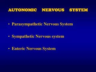
Lecture 9: Autonomic Nervous System
320 likes | 803 Vues
Lecture 9: Autonomic Nervous System. Controls the function of internal organs. The autonomic nervous system provides internal homeostasis. Autonomic reflexes control blood pressure, heart rate, respiration, water balance, body temperature and other homeostatic functions.

Lecture 9: Autonomic Nervous System
E N D
Presentation Transcript
Controls the function of internal organs. • The autonomic nervous system provides internal homeostasis. • Autonomic reflexes control blood pressure, heart rate, respiration, water balance, body temperature and other homeostatic functions. • Divided into two major subdivisions: the sympathetic and parasympathetic divisions. The two divisions cannot be readily distinguished except according to the type of situation in which they are most active.
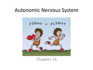
Resulting Homeostasis Following a Balance Between Sympathetic and Parasympathetic Control F11-1 The fight-or-flight response is the quick protective reaction of the body
Two Neurons Form Synapses in Ganglia • Ganglia can modulate information passing through them with the assistance of intrinsic neurons. • A single preganglionic neuron synapses with as many as 8-9 postganglionic neurons. Allows control of multiple pathways.
Neuroeffector Junction F11-3 • Varicosities release neurotransmitters. Lay close to targets. • Diffused transmitter interacts with receptors, wherever they are. Less directed communication, but more tissue affected.
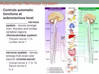
Sympathetic Pathway • Originate from the thoracic and sacral regions of the spinal cord. • Ganglia are found in the sympathetic chain. • Postganglionic neurons have long axons. Parasympathetic Pathway • Preganglionic cell bodies are found in the brain stem or the sacral region of the spinal cord. • Parasympathetic ganglia are found near their target organ. • Preganglionic axons are long.
Vagus Nerve • Carry sensory neurons and parasympathetic fibres to most internal organs. • Vagotomy- performed in the late 19th and early 20th centuries to treat stomach ulcers by removing the parasympathetic innervation which causes acid secretion. Nowadays, its treated with drugs. • Otto Loewi: Discovered chemical synaptic transmission by showing that the slowing of the heart beat upon vagal nerve stimulation resulted from the release of ACh from nerve endings. The details of his important experiment came to him in his dream. He won the Nobel Prize in 1936.
Adrenal Medulla Secretes Catecholamines • The adrenal medulla is a modified sympathetic ganglion. Postganglionic neurons lack axons. • NE and E are secteted in response to alarm signals from the CNS into the bloodstream for distribution throughout the body.
ACh and NE are the Primary Neurotransmitters • Neurons which secrete ACh are termed cholinergic and those which secrete NE are termed adrenergic.
Adrenergic neurons are mainly sympathetic. • All preganglionic neurons are cholinergic. Parasymapthetic neurons release ACh at neuroeffector synapses. • A few sympathetic neurons release ACh instead of NE and they are termed sympathetic cholinergic neurons. • Non-adrenergic, non-cholinergic neurons • They secrete neither ACh or NE. They can also secrete transmitters such as VIP, substance P, NO, ATP, along with ACh and NE. • They are assigned to either autonomic division according to their anatomical organization. • Geoffrey Burnstock formulated the concept of purinergic signaling. ATP is a purine nucleotide. It is a neurotransmitter in the ANS; functions by activating ‘purinoceptors’.
Neurotransmitter Release F11-8 • Neurotransmitters are synthesized and packaged into vesicles within the varicosities. • Presynaptic receptors can modulate release. Facilitate or inhibit it. • Substances are co-released along with transmitters.
Termination of Neurotransmitter Release • Neurotransmitters can be metabolized in the extracellular fluid by enzymes (eg. ACh by AChE). • Transported back into the axon terminal (eg.NE). • The concentration of neurotransmitter in the synaptic cleft and the number of receptors on target cells determine the magnitude of the synaptic response.
Cholinergic Neurotransmitter Receptors F11-7 • nAChR open cation channels which pass Na+ and K+. Net Na+ ion influx which depolarizes postsynaptic cell. • mAChR are G-protein coupled channels linked to 2nd messengers or gated to K+ channels. The tissue response varies with receptor subtype.
Adrenergic Receptors • Adrenergic receptor activates responses which lasts for long periods resulting from 2nd messenger activation. It modifies protein synthesis or existing proteins. • Three subtypes: a, b1and b2.
a receptors- Cause muscle contraction. In the digestive tract, they cause relaxation. • b1receptors- enhance cardiac contraction and speed up electrical conduction in cardiac muscle. • b2 receptors- activation by epinephrine triggers smooth muscle relaxation.
Beta-blockers are used clinically to treat high blood pressure; one of the most common disorders in the US.
Homeostatic Regulation by the Autonomic Divisions • The two autonomic divisions demonstrate all properties of homeostasis: • 1) Maintenance of the internal environment. • 2) “Up-down” regulation by varying the tonic control. • 3) Antagonistic control. • 4) Variable tissue responses that result from different membrane receptors. Eg. blood vessels which contain receptors which mediate vasoconstriction, whereas another class of receptors cause vasodilation. • Eg. Sympathetic stimulation increases the heart rate whereas parasympathetic stimulation decreases it. Thus, the heart rate could be altered by changing the relative contribution of the two divisions. Example displays (2), (3) and (4).
Autonomic Control is Regulated by the Brain F11-9 • CNS control of autonomic divisions is closely linked to centres in the medulla, pons and hypothalamus.
Hypothalamus Integrates Many Autonomic Reflexes F9-15 • Controls homeostasis and behavioural drives. • Receives input from the limbic system. • Best known hypothalamic response is the fight-or-flight response. It increases sympathetic output: pounding heart, sweaty palms and increased blood pressure in response to fear. • Catecholamine release from the adrenal medulla spreads this response far and wide through the bloodstream.
References Tortora, G.J. & Grabowski, S.R (2003). Principles of Anatomy & Physiology.New Jersey: John Wiley & Sons. Ch.17, pp.565-581. Silverthorn, D.U (1998). Human Physiology: An Integrated Approach. New Jersey: Prentice Hall. Ch.11, pp.307-318.