Download
1 / 5
50 likes | 418 Vues
BIO 210 Handout #5A – Epithelial, Muscle and Nervous Tissues. SHAPES. ARRANGEMENTS. Cell – basic structural and functional unit of living organisms Tissue – groups of cells that are similar in structure and function Histology – study of tissues Four primary tissue types – Epithelium –

E N D
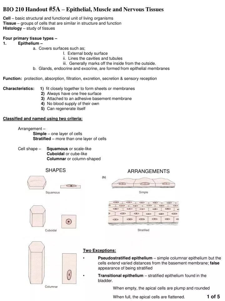
BIO 210 Handout #5A – Epithelial, Muscle and Nervous Tissues SHAPES ARRANGEMENTS • Cell – basic structural and functional unit of living organisms • Tissue – groups of cells that are similar in structure and function • Histology – study of tissues • Four primary tissue types – • Epithelium – • a. Covers surfaces such as; • I. External body surface • ii. Lines the cavities and tubules • iii. Generally marks off the inside from the outside. • b. Glands, endocrine and exocrine, are formed from epithelial membranes • Function: protection, absorption, filtration, excretion, secretion & sensory reception • Characteristics: 1) fit closely together to form sheets or membranes • 2) Always have one free surface • 3) Attached to an adhesive basement membrane • 4) No blood supply of their own • 5) Can regenerate itself • Classified and named using two criteria: • Arrangement – • Simple – one layer of cells • Stratified – more than one layer of cells • Cell shape – Squamous or scale-like • Cuboidal or cube-like • Columnar or column-shaped • Two Exceptions: • Pseudostratified epithelium – simple columnar epithelium but the cells extend varied distances from the basement membrane; false appearance of being stratified • Transitional epithelium – stratified epithelium found in the bladder. • When empty, the apical cells are plump and rounded • When full, the apical cells are flattened. 1 of 5
Epidermis Esophagus Simple Squamous Epithelia – single layer of flattened cells Location: Air sacs of lungs, lining of heart, blood vessels, lymphatic vessels, ventral cavity Lung air sac Stratified Squamous Epithelium – many cell layers. Basal cells are cuboidal, surface cells are flattened Location: moist linings of esophagus, mouth, vagina. Keratinized – skin epidermis Simple Cuboidal Epithelium – single layer of cube-like cells, large central nucleus Location: lining the digestive tract, gallbladder, ducts of some glands Ciliated – small bronchi, uterine tubes, some parts of uterus 2 OF 5
Simple Columnar Epithelium – single layer of tall cells May be ciliated and/or contain Goblet cells Location: lining digestive tract, gallbladder, ducts of some glands Ciliated – small bronchi, uterine tubes, some parts of uterus Trachea Pseudostratified Columnar Epithelium – single layer of cells of differing heights Nuclei seen at different levels; may contain Goblet cells Location: ducts of large glands, part of male urethra Ciliated – trachea, most of upper respiratory tract Transitional Epithelium – resembles both stratified squamous & stratified cuboidal Surface cells may be dome-shaped or flattened Location: bladder, ureters, part of the urethra 3 OF 5
Muscle Tissue – highly specialized to contract in order to produce movement of some body parts. Cells are quite elongated to provide a long axis for contraction. Three basic types: 1. Skeletal – long, cylindrical, multinucleate cells; obvious striations (bands attached to the skeleton. It is under voluntary control. Function: Contractions move the limbs and other external body parts 2. Cardiac Muscle – found only in the heart; Cardiac cells are uninucleate. Branch & connect to other cardiac muscle cells at junctions called intercalated discs. It is under involuntary control. Function: when contraction occurs, blood is propelled through the blood vessels 3. Smooth Muscle – visceral muscle, Spindle shaped cells with central nucleus, no striationsforms sheets;found mainly in the walls of hollow organs (digestive & urinary tract organs, uterus, blood vessels). They are under involuntary control. Function: Contraction constricts or dilates the lumen and propels substances forward. 4 of 5
Nervous Tissue – two major cell populations Neuroglia – special supporting cells that protect, support and insulate delicate neurons Neurons – highly specialized with a nucleus-containing cell body and their cytoplasm drawn out into long extensions. Found in the brain, spinal cord, nerves Function: - transmit nerve impulses from sensory receptors to effectors (muscles & glands) Neuron Identify the following tissues: A. B. D. _____________________ C. Identify the tissue lining the lumen of the vein E. ____________________ F._____________________________ 5 of 5