Download

1 / 55
570 likes | 811 Vues
Forensic Biology Screening Workshop Other Body Fluids and Tissues. Vaginal Secretions. Vaginal Secretions. Vaginal secretions are a complex mixture of cells and secretions There is no confirmatory test to identify vaginal secretions

E N D
Forensic Biology Screening WorkshopOther Body Fluids and Tissues
Vaginal Secretions • Vaginal secretions are a complex mixture of cells and secretions • There is no confirmatory test to identify vaginal secretions • Several screening tests based on microscopy have been proposed.
Vaginal Secretions • Vaginal epithelial cells are large, and many contain glycogen (a polysaccharide) which can be demonstrated by staining with iodine in the form of a solution or exposing to iodine vapor. • The presence of glycogenated cells is variable depending on the stage of the estrous cycle • Glycogenated cells can be found in other body secretions (i.e. oral, anal)
Vaginal Secretions • Based on the very high glycogen content of the vagina, vaginal secretions have a higher quantity of glycogenated cells than other body fluids • Lugol's solution, first made in 1829, is a solution of iodine • Named after French doctor, Lugol
Lugol’s Staining • Squamous epithelial cells with high glycogen content will show a chocolate brown color in the body of the cell (cytoplasm) with a clear round to oval shaped unstained nucleus located somewhat centrally in the cytoplasm
Lugol’s Staining HOW TO MAKE: • Iodine (0.02m) 1 g • Potassium iodide (0.06m) 2 g • Distilled water 200 ml Dissolve the potassium iodide in the water, then add iodine. HOW TO STORE: • Relatively stable at room temp or 4C
Lugol’s Staining HOW TO PERFORM: • Make a thin smear of the questioned material on a glass microscope slide • Fix the smear by gentle heating • Cover the smear with Lugol’s iodine solution • Add an equal volume of water to the smear
Lugol’s Staining HOW TO PERFORM: • Place a cover slip on the stained area • Allow to stand 3-5 minutes at room temperature • Observe microscopically at 200-400x
Lugol’s Staining HOW TO PERFORM: • Squamous epithelial cells with high glycogen content (notably vaginal and penile urethral epithelial cells) will exhibit a chocolate-brown or tan color • Other epithelial cells will exhibit a yellow or gold color
Lugol’s Staining HOW TO PERFORM: • A majority of epithelial cells stained chocolate brown is a positive result. • Known vaginal cells should be tested as a positive control • Known buccal cells should be tested as a negative control
Lugol’s Staining epithelial cells stained chocolate brown • Gold/yellow stained epithelial cells
Fecal Material • Feces are food residues passed after completion of travel through the digestive system • Has a characteristic odor mainly due to skatole, an organic compound that occurs naturally in feces
Fecal Material Microscopy • Microscopy has been used to identify fecal material • Looking for undigested residues of food material Chemical Tests • Detection of urobilinogen, a bile pigment excreted in feces, which may be detected using its fluorescent reaction to Edelman’s reagent
Test for Urobilinogen HOW TO MAKE: • Solution #1: 40% Alcoholic mercuric chloride solution • Mercuric chloride 4 g • Methanol 10 ml Mix and store in stopper bottle • Solution #2: 40% Alcoholic zinc chloride solution • Zinc chloride 4 g • Methanol 10 ml Mix and store in stopper bottle • Solution #3: Amyl alcohol
Test for Urobilinogen HOW TO STORE: • Relatively stable at room temp or 4C
Test for Urobilinogen HOW TO PERFORM: • Extract the suspected stain and an unstained control in 3 drops of water in separate test tubes • Place an equal amount of water in a third test tube for the negative control • Extract a known stain in 3 drops of water • Add 3 drops of 40% alcoholic mercuric chloride to each tube
Test for Urobilinogen HOW TO PERFORM: • Add 3 drops of amyl alcohol to each tube. Shake to mix • Centrifuge and collect the clear supernatant • Examine under UV long-wave light for any fluorescence No fluorescence should be visible at this point in the analysis • Add 3 drops of 40% alcoholic zinc chloride to each tube and shake
Test for Urobilinogen HOW TO PERFORM: • Incubate at room temperature for 30-60 minutes • Examine under long-wave UV light. The appearance of a green fluorescence is considered a positive test for urobilinogen, indicative of the presence of fecal matter
Test for Urobilinogen HOW TO PERFORM: • Known fecal material should be tested as a positive control • Water should be tested as a negative control Note: Safety eyeglasses which absorb ultraviolet radiation must be worn when examining for fluorescence with UV light
Urine • No confirmatory tests for the presence of urine • Urine stains fluorescent under ultraviolet light • This can be useful for locating stains prior to chemical testing • Has a characteristic odor • Contains a large amount of urea, a chemical byproduct of normal metabolic processes in the body • Identification of high levels of urea can serve as a screening test for urine in fluids or stains • Perspiration can give reactions similar to urine
Urine • Contains creatinine, which is a breakdown product of creatine (an important part of muscle) • Over time, the creatine molecule gradually degrades to creatinine • Creatinine is a waste product that is excreted from the body entirely by the kidneys • Identification of creatinine can serve as a screening test for urine in fluids or stains
Urea-Litmus Paper Test Litmus Paper test for the detection of ammonia • Relies on the indirect identification of urea by reacting a test sample with urease to generate ammonia from the urea. Litmus paper is used to detect the ammonia Urea + H2O ↔ CO2 + 2NH3 • A known urine sample and a blank are tested as a positive and negative control. A substrate control may be tested, as needed
Urea-Litmus Paper Test • Affix a strip of litmus paper to a cork or rubber stopper that will fit into the test tubes used: cut a slit in the bottom of the stopper and insert one end of the paper. The paper strip should hang down into the tube without touching the sides of the tube or becoming wet • Place a cutting from a suspected urine stain in a test tube with 3-4 drops of distilled water • Add a drop of urease solution (urease mixed with distilled water) to each tube
Urea-Litmus Paper Test • Incubate at 37°C for 30 minutes • A definite and easily observed color change to blue on the litmus paper represents a positive result
Urea-Nitrogen Tube Test • Relies on the indirect identification of urea by reacting a test sample with urease to generate ammonia from the urea. Ammonia is subsequently identified by the production of a deep blue-colored solution Urea + H2O ↔ CO2 + 2NH3
Urea-Nitrogen Tube Test HOW TO MAKE: • A urease buffer solution • A urea-nitrogen standard solution • A phenol-nitroprusside reagent • An alkaline hypochlorite reagent
Urea-Nitrogen Tube Test HOW TO STORE: • The urease buffer solution is not stable at 4° C. for more than a few weeks. Make fresh solution according to concentration stated on label (30 mg urea/100 ml distilled water) • Other reagents-see product inserts
Urea-Nitrogen Tube Test • A known urine sample and a blank are tested as a positive and negative control. A substrate control may be tested, as needed
Urea-Nitrogen Tube Test Step 1
Urea-Nitrogen Tube Test • Cover tubes and allow to stand 20 minutes at room temperature • Add 1.0 ml phenol-nitroprusside reagent to each tube • Add 1.0 ml alkaline hypochlorite reagent to each tube • Invert to mix. Read results within 5 minutes.
Urea-Nitrogen Tube Test Question Sample • If stain is urine, a deep blue color should be observed which should be as intense as the urine positive control. • If the color is not as intense as the urine positive control or the color in the question sample (without urease) tube is as intense as the sample tube, this constitutes an inconclusive test result • If no color is observed, this is a negative result
Creatinine Jaffe Test • One of the oldest tests for the detection of creatinine-1886 • Creatinine forms a red compound with picric acid (Jaffe test)
Jaffe Test HOW TO MAKE: • Saturated aqueous picric acid solution • 1 gram picric acid in 30 ml H2O • 5% Sodium hydroxide • Glacial acetic acid
Jaffe Test HOW TO PERFORM: • Cut a portion of the suspected stain and an unstained control area and place on filter paper • Cut a portion of a known urine stain and place on a separate filter paper • To each, add 1 drop of saturated picric acid solution followed by 1 drop of 5% NaOH
Jaffe Test • An orange coloration after 15 minutes on the questioned stain and surrounding filter paper is considered a positive result. The control area should remain yellow. • Addition of 2 drops of glacial acetic acid should render the orange questioned stain yellow (de-colorization), further confirming the presence of creatinine
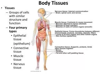
Hair • Composed of cylindrical structures or shafts made up of tightly compacted cells that grow from follicles • Diameter ranges from 15-120 µm • Depends on type of hair and body region • Root material can be used for nuclear DNA testing • Shaft material can be used for mtDNA testing
Hair • Human hairs are distinguishable from hairs of other mammals • Human hairs are generally consistent in color and pigmentation throughout the length of the hair shaft, whereas animal hairs may exhibit radical color changes in a short distance, called banding • The medulla, when present in human hairs, is amorphous in appearance, and the width is generally less than one-third the overall diameter of the hair shaft. The medulla in animal hairs is normally continuous and structured and generally occupies an area of greater than one-third the overall diameter of the hair shaft.
Hair Structure • Three cell types • Outer cuticle • Central cortex • Central medulla
Hair • In some instances human hairs can be classified by racial origin such as Caucasian (European origin), Negroid (African origin), and Mongoloid (Asian origin). • In some instances the region of the body where a hair originated can be determined by its gross appearance and microscopic characteristics • The length and color can be determined • It can also be determined whether the hair was forcibly removed, damaged by burning or crushing, or artificially treated by dyeing or bleaching.
Hair The growth phase of the hair is important in determining whether the root is suitable for nuclear DNA analysis testing • Growth Cycles • Anagen phase • Catagen phase • Telogen phase
Hair Anagen Phase • Active hair growth • Contains nucleated cells in the root and in the surrounding sheath material • Generally suitable for nuclear DNA analysis
Hair Anagen Hair
Hair Catagen Phase • Transitional phase after active hair growth, cell division stops • Characteristic club appearance of root • May be suitable for nuclear DNA analysis
Hair Catagen Hair
Hair Telogen Phase • Follows transitional phase-growth ceases • Shedding phase • Telogen hairs without follicular tissue may not be amenable to nuclear DNA analysis because of the lack of nucleated cells • May contain sufficient mitochondrial DNA in their roots and hair shafts for analysis