Download
1 / 56
670 likes | 1.34k Vues
Physiology of blood system. Blood system. Blood system firstly was proposed by Lung in 1936. It consist of - blood circulated through the blood circulatory system - blood forming organs - blood destroying organs - regulatory apparatus. Blood.

E N D
Blood system • Blood system firstly was proposed by Lung in 1936. • It consist of • - blood circulated through the blood circulatory system • - blood forming organs • - blood destroying organs • - regulatory apparatus.
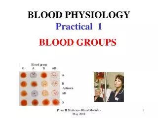
Blood • Blood is a fluid connective tissue. Blood consist of • - plasma • - blood cells – erythrocytes, • leucocytes and • platelets.
plasma Leucocytes and platelets erythrocytes
Amount of blood • The amount of blood in the body has been measured in various ways. Naturally the volume of blood can be expected to vary with the size of the body. The blood volume of an adult human of average size is about 6-8 % (in man – 52-83 mL/kg; woman – 50-75 mL/kg).
Plasma • Water – 90% • Solids – 10% • Inorganic chemicals: sodium, calcium, potassium, magnesium, chloride, bicarbonate, phosphate, sulfate – 0,9 % • Organic chemicals: • Proteins: serum albumin, serum globulin, fibrinogen – 8 % • Others: – 1,1 % • Nonprotein nitrogenous substances: urea, uric acid, creatine, creatinine, ammonium salts, amino acids • Nonnitrogenous substances: glucose, fats, cholesterol hormones • Gases: oxygen, carbon dioxide, nitrogen
Proteins • One liter of plasma has 65-85 gram of proteins. • Concentration of albumins is 35-50 g/L; globulins is alpha-1-globulins – 1-4 g/L, • alpha-2-globulins – 4-8 g/L, • beta-globulins – 6-12 g/L, • gamma-globulins – 8-16 g/L; • fibrinogen – 2-4 g/L. • Plasma which are not contain fibrinogen called serum (it is necessary for understanding the immunology, therapy etc.)
Albumins • Albumins: on 80 % it provides oncotic pressure, contacts with bilirubin, fat acids, antibiotics, sulfanilamids. It connects with them and transports them. It produces in liver in average quantity of 17 gram per day.
Globulins • Globulins produces in lymphatic nodes, in liver, in bone marrow in average quantity of 5 gram per day. • Alpha-1-globulins connected with carbohydrates (for example 2/3 of all glucose connected with alpha-1-globulins. This is glyco proteins.) • Alpha-2-globulins connect 90 % of cupper. This is cerruloplasmin. Its may produced in hormons, for example, thyroxin, connected by vitamin B12. From this protein produce angiotensines (substances which are take place in increase of blood pressure). • Beta-globulin carry out 75 % of fats, iron (for example, transferrine). • Gamma-globulins has protective functions (for example, antibodies).
Fibrinogen • Fibrinogen is a protein which are produced by liver and take place in hemostasis system. Fibrinogen is dissolved form, which transform in insolved form – fibrin and provide coagulative hemostasis (plug production) and prevent bleeding. • Daily production of fibrinogen is 2-4 g/L.
Quantity of cells, their changing • Erythrocytes (In men – 4,0-5,1 Tera/L; in women – 3,7-4,7 Tera/L. The quantity of erythrocytes may be increase – in pregnancy, in physical training, mental work, in newborn or decrease.) • Leukocytes (Their number are 4-9 Giga/L. The number of leukocytes may increase – physical work, emotional load, in newborn, inflammation or decrease.) • Platelates (Their number are180-320 Giga/L.)
Functions of blood • 1. Breathing function of blood. • 2.Trophic function of blood. • 3.Excretory function of blood. • 4.Hormonal regulation. • 6. Temperature regulation. • 7.Maintaining the acid-base balance of tissues. • 8.Supporting the water-electrolytic balance. • 9.Homeostasis function. • 10.Protecting the body from bacteria and other organisms that can cause diseases or other abnormal conditions.
Respiratory pigments • HemoglobinErythrocytes derive their colour from a complex protein called hemoglobin. This substance is composed of a pigment, heme, containing iron, and the protein glohin. Hemoglobin has the power to attract oxygen molecules and to hold them in a loose chemical combination known as oxyhemoglobin. It is said, therefore, to have a chemical affinity for oxygen.
Respiratory pigments • MyoglobinHem is also part of the structure of myoglobin, an oxygen-binding pigment found in red (slow) muscles and in the respiratory enzyme cytochrome c. Porphyrins other than that found in hem play a role in the pathogenesis of a number of metabolic diseases (congenital and acquired porphyria, etc.) It may be the reserve pigments, which give the tissue oxygen in a small oxygen condition.
Exchange of iron in the organism • In the blood-destroying organs, the hemoglobin breaks down into an iron-free and the iron-bearing portions. The latter is decomposed into bilirubin and an iron compound. Both are carried to the liver, where the bilirubin is excreted in the bile as one of the bile pigments, while the iron, if not needed for the formation of new red blood cells, is stored. Other way entering of iron is the food. Erythrocytes can live only a limited time. The life of red blood cells are nearly 120 days. Blood cells are lost by the processes of hemolysis and fragmentation, which occur throughout the circulatory system, and phagocytosis of whole cells and cell fragments, which takes place in the cells of the reticuloendothelian tissues, especially those in the spleen, the liver, and the bone marrow.
Function of leukocytes • 1. Protective • 2. Transport • 3. Metabolic • 4. Regenerator
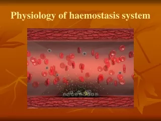
Hemostasis system • Hemostasis is very important for our life, because if we are live our hemostatic system is very strong. They are includes in a case of trauma, cutting the vessels etc. Hemostasis isthe physiologic system, which support the blood in the fluid condition and prevent bloodless. Hemostasis system vital necessary and functionally connect with the cardiovascular, breathing, endocrine and other systems.
The components of hemostasis • The components of hemostasis are wall of the vessels, blood cells – platelets, erythrocytes, leucocytes, enzymes and nonenzymes components of plasma – clotting and anticlotting substances, fibrinolysis components of hemostasis. There are 2 kinds of hemostasis. They are vessel-platelets (primary) and coagulative (secondary) hemostasis. Primary hemostasis activity begin the first after the destroyed of vessels. Secondary hemostasis starts after that in case the primary hemostasis do not stopped the bloodless.
Vessel-platelets hemostasis (or primary hemostasis include in clotting first of all after the destroyed the safe of vessel wall.) • Properties and function of platelets • Quantity of platelets is 180-320 G/L. Diameter of platelets is 1-4 micrometers, thickness – 0,5-0,75 micrometers. They are the little peace of megacariocytes cytoplasm (from one megacariocytes may develop few hundred of platelets). Platelets circulated in blood from 5 to 11 days and than destroyed in liver, lungs, spleen by the cells of macrophagal system.
Function of platelets are: • 1. hemostatic function – platelets produce substances, which are secure the hemostasis. 2. Angiotrophic function – provide trophic of endotheliocytes of vessel wall, support structure and functions of microvessels. These function is realize by adgesion of platelets to endotheliocytes and injection the enzymes into the endotheliocytes. For one day near 35 G/L platelets do this function. 3. Transport function – transfer the enzymes, ADP, serotonin and other. 4. Phagocytosis function – the contain of platelets help to kill viruses and antigens bodies. 5. Regeneratory function – platelets have the growth factor, which help to grow the endothelial and muscles cells which are present in the vessel wall.
Stages of vessel-platelets hemostasis • 1. Shorting spasm of the vessels – vascular spasm duration to 1 minute is caused by catecholamins and other enzymes. Diameter of vessels decrease on ½-⅓. Mechanism of it development determine by secretion of serotonin and thromboxan A2 from platelets and epinephrine from ending of sympathetic nerves. • 2. Adgesion of platelets – activation of platelets and stick it to the place of defect in vessel wall. • 3. Reverse aggregation of platelets – the thromb which are formed may make way for plasma. • 4. Unreverse aggregation of platelets – the thromb which are formed can not may make way for plasma. • 5. Retraction of platelets plug – decrease the size of plug, pack down the plug.
Investigation of vessel-platelets hemostasis • 1. Calculation of the platelets quantity 180-320 G/L. • 2. Determination of duration of capillary bleeding after Duke’s method – to 3 minute in norm. • 3. Sample of fragility of capillars – to 10 petechias in norm in a round with diameter 5 santimetres.
Coagulative (secondary) hemostasis. • Characteristics of clotting factors • There are 12 clotting factors: • I – fibrinogen; II – prothrombine; III – thromboplastin of tissue; IV – ions of calcium; V – proaccelerin; VII – proconvertin; VIII – antihemophylic factor A; IX – Christmas factor or antihemofilic factor B; X – Stuart-Prower factor or prothrombinase; XI – plasma thromboplastin antecedent; XII – Hageman factor; XIII – fibrin stabilizing factor.
Some of them are enzymes – II, VII, IX, X, XI, XII,XIII; otherare not – I, III, IV, V, VIII. The vitamin K is necessary for the functional activity of II, VII, IX, X factors.
External mechanism of the first stage (3 factors from the injure tissues go to plasma and interactions with VII factor, the last is activated. VII active factor and IV factors form the complex 1a: III + VII active + IV, which is activated X factor.)
Inner mechanism of the first stage (Factor 3 of platelets – platelets thromboplastine – influence on XII factor. Active XII factor + XI is complex 1. Active XI factor activated IX factor. Active IX factor + VIII factor + IV factor is complex 2. Complex 1a and 2 are activate X factor. Factor X active + V + IV formed complex 3 or thrombinasa complex.) • Course of the second and third stages (The second stage – formation of thrombin from prothrombin. The third stage is formation of fibrin from fibrinogen. The last stage has 3 period; formation of fibrin-monomers; formation of fibrin S (solubilis); formation of fibrin I (insolubilis). Calcium is necessary for all stages.)
Regulation of the clotting mechanisms • Increase of clotting names hypercoagulation, decrease – hypocoagulation. Hypercoagulation may be in a stress cases. It depends on epinephrine, which concentration increased in the cases of stress. Epinephrine increase from the vessels walls factors from which produced prothrombinasa. In cases of big concentration epinephrine should activate XII factor in a bloodstream. It divides fats and fat acids, which have prothrombinase activity. After the hypercoagulation stage may be secondary hypocoagulation.
Coagulogram • Time of clotting by Ly-Wait – 5-10 minutes; time of plasma recalcification – 60-120 seconds; thrombotest – IV, V, VI degree; thromboplastin time – 12-15 seconds; thromboplastin index – 80-105 %; concentration of fibrinogen – 2-4 g/L; tolerancy of plasma to heparin – 6-11 minutes; heparin time – 50-60 seconds; fibrinolysis – 15-20 %.