Microbial Growth
Microbial Growth. Microbial growth implies an increase in cellular constituents - leads to rise in cell number when microorganisms reproduce by processes like budding or binary fission - ability to reproduce is a major criterion to determine if

Microbial Growth
E N D
Presentation Transcript
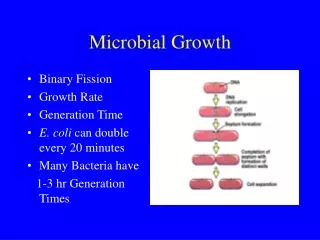
Microbial Growth Microbial growth implies an increase in cellular constituents - leads to rise in cell number when microorganisms reproduce by processes like budding or binary fission - ability to reproduce is a major criterion to determine if a microbe is alive or not - results when cell become longer/larger Population growth is used to analyze the growth curve of a microbial culture
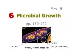
Cell Growth and Binary Fission Fts proteins forms in the space between the duplicated nucleoids Min E proteins assist in the location of the actual cell midpoint
forms a ring around the cylinder in the center of the cell The FtsZ ring and cell division
2 copies of chromosomes are pulled apart to each daughter cell
Peptidoglycan Features 1. 3-D polymeric macromolecule 2. Formed from subunits by two types of covalent bonds 3. ß-1,4 glycosidic bonds between hexose sugars, and peptide bonds between amino acids 4. Determines cell shape and prevents osmolysis 5. Dynamic structure a) must grow as cell grows b) must be regulated to allow septation
Peptidoglycan Glycan chains MA -1,4-linkages L-ala GA D-glu MA MA L-lys Tetrapeptide L-ala L-ala GA D-ala GA D-glu D-glu MA L-lys L-lys L-ala GA D-ala D-ala D-glu Peptidecross-link L-lys D-ala
Gram - negative Gram - positive
Peptidoglycan-Targeting Antibiotics • Destruction of peptidoglycan causes bacterial lysis • This can be accomplished in the laboratory using the enzyme lysozyme, which hydrolyzes the glycosidic linkages • Antibiotics should target bacteria-specific processes, such as peptidoglycan synthesis • DO NOT use these antibiotics on bacteria with no cell wall (Mycoplasma)or a cell wall that is not susceptible to them (Mycobacteria)
Antibiotics that Target the Peptidoglycan • Phosphonomycin • Cycloserine • Vancomycin • Bacitracin • Penicillin • Cephalosporins
Synthesis-Cytoplasm D-ala-D-ala added as a dipeptide Start NAG converted to NAM L-ala, D-glu and L-lys added one at a time
Synthesis-Membrane Addition of NAG results in dipeptide precursor Start Transfer to lipid carrier
Sites of action of different antimicrobial agents. PABA, paraminobenzoic acid; DHFA, dihydrofolic acid; THFA, tetrahydrofolic acid.
Outer wall of Gram-positive and Gram-negative species and detail of porin channels of Gram-negative bacteria. Antimicrobial agents diffuse easily through the loose outer wall of Gram-positive bacteria, but must go through the narrow channels of the Gram-negative species.
Structure of metronidazole and its mechanism of action. Metronidazole enters an aerobic bacterium where, via the electron transport protein ferrodoxin, it is reduced. The drug then binds to DNA, and DNA breakage occurs.
Diagrammatic representation of inhibition sites of protein biosynthesis by various antibiotics that bind to the 30S and 50S ribosomes.
Structure of sulfonamide and trimethoprim with sites of inhibition of folic metabolism.
MULTIDRUG RESISTANCE AMONG PATHOGENIC BACTERIA How do they do it? • Acquire ability to degrade the antibiotic • Change their outer structure to prevent drug entry • Change the drug target so that it is no longer affected by the drug • Acquire and/or turn on an efflux pump to eliminate the drug from the cell
Where do they get it? • Chromosomal mutation(s) under selective pressure by the antimicrobial • Conjugal transfer of resistance plasmids • Conjugal transfer of chromosomal resistance genes • Infection by bacteriophage • An old system that found a new use (efflux pumps)
Example of how two antibiotics (A and B) may interact with synergy, indifference, or antagonism.
Types of Antibiotics Penicillins have a common chemical structure which they share with the cephalopsorins. Penicillins are generally bactericidal, inhibiting formation of the cell wall. Types of penicillin • The natural penicillins are based on the original penicillin-G structure. • Penicillin-G types are effective against gram-positive strains of streptococci, • staphylococci, and some gram-negative bacteria such as meningococcus. • Penicillinase-resistant penicillins, notably methicillin and oxacillin, are active • even in the presence of the bacterial enzyme that inactivates most natural • penicillins. • Aminopenicillins such as ampicillin and amoxicillin have an extended spectrum • of action compared with the natural penicillins. Extended spectrum penicillins • are effective against a wider range of bacteria.
Penicillin • Penicillin binds to proteins known as Penicillin-Binding Proteins (PBPs) • Multiple PBPs are made by each species, with different molecular weights and different enzymatic activities • PBPs are involved in the cross-linking reactions, and typically have transpeptidase activity • Inhibition of peptidoglycan cross-linking destabilizes the cell wall
Cephalosporins Cephalosporins have a mechanism of action identical to that of the penicillins. However, the basic chemical structure of the penicillins and cephalosporins differs in other respects, resulting in some difference in the spectrum of antibacterial activity. Like the penicillins, cephalosporins have a beta-lactam ring structure that interferes with synthesis of the bacterial cell wall and so are bactericidal. Cephalosporins are derived from cephalosporin C which is produced from Cephalosporium acremonium.
Tetracycline Tetracyclines got their name because they share a chemical structure that has four rings. They are derived from a species of Streptomyces bacteria. Tetracycline antibiotics are broad-spectrum bacteriostatic agents, that inhibit bacterial protein synthesis. Tetracyclines may be effective against a wide variety of microorganisms, including rickettsia and amebic parasites. Structure of tetracycline showing the area critical for activity and major and minor points of modification.
Macrolides The macrolide antibiotics are derived from Streptomyces bacteria, and got their name because they all have a macrocyclic lactone chemical structure. The macrolides are bacteriostatic, binding with bacterial ribosomes to inhibit protein synthesis. Erythromycin, the prototype of this class, has a spectrum and use similar to penicillin. The most commonly prescribed macrolide antibiotics are: erythromycin clarithromycin azithromycin dirithromycin roxithromycin troleandomycin
Fluoroquinolones Fluoroquinolones (fluoridated quinolones) are the newest class of antibiotics. Their generic name often contains the root "floxacin". They are synthetic antibiotics, and not derived from bacteria. Fluoroquinolones belong to the family of antibiotics called quinolones. The older quinolones are not well absorbed and are used to treat mostly urinary tract infections. The newer fluroquinolones are broad-spectrum bacteriocidal drugs that are chemically unrelated to the penicillins or the cephaloprosins. Because of their excellent absorption fluroquinolones can be administered not only by intravenous but orally as well.
Aminoglycosides Aminoglycoside antibiotics are used to treat infections caused by gram-negative bacteria. Aminoglycosides may be used along with penicillins or cephalosporins to give a two-pronged attack on the bacteria. The aminoglycosides are drugs which stop bacteria from making proteins. This effect is bacteriocidal. The most commonly-prescribed aminoglycosides: amikacin gentamicin kanamycin neomycin streptomycin tobramycin
MICROBIAL GROWTH KINETICS • - describe how the microbe grows in the fermenter • - important to determine optimal batch times • growth of microbes can be broken down into 4 stages; • LAG PHASE • LOG/EXPONENTIAL PHASE • STATIONARY PHASE • DEATH PHASE accelerated death phase decelerated growth phase Lag Phase Cells have just been introduced into a new environment Cell growth is minimal Cell is synthesizing new components – no cell division takes place accelerated growth phase • cell is old and depleted of ATP • medium may be different from the one the microorganism was growing • microorganism have been injured and require time to recover
LOG/EXPONENTIAL PHASE Cells have adjusted to their environment Rapid growth takes place Cell growth rate is highest in this phase At some point, cells growth rate level off and become constant STATIONARY PHASE Cell growth rate has leveled off and become constant Number of cells multiplying equals the number of cells dying - nutrient limitation - aerobic organism are limited by oxygen availability - population growth cease due to the accumulation of toxic waste products DEATH PHASE Decline in the number of viable cells
Log/Exponential Phase The rate of increase in biomass is correlated with the specific Growth rate µ and the biomass concentration X (g/L), whereas the Rate of sincrease in cell number is correlated with µ and cell density N (1/L) d X = µ • X or d N = µ • N d t d t specific growth rate, µ, 3 parameters : the concentration of limiting substrate S the maximum growth rate µmv the substrate-specific constant Ks µ = µm ___S___ Ks + S Monod equation Ks substrate concentration at which half the maximum specific growth rate is obtained (µ = 0.5 µm ) - equivalent to the Michaelis constant in enzyme kinetics
Maximal specific growth rates (µm) of some fungi on glucose Anderson et al, 1975
Effect of glucose chain length on the maximal specific growth rate (µm) in Fusarium graminearum at 30 ºC Anderson et al, 1975
PARAMETERS THAT MUST BE PRECISELY REGULATED • temperature • pH • rate and nature of mixing • oxygenation • sterility and containment Types of Cultures 1. Batch 2. Fed-batch 3. Continuous 4. Synchronous