Download
1 / 24
240 likes | 502 Vues
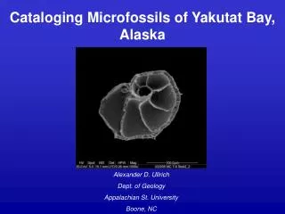
Cataloging Microfossils of Yakutat Bay, Alaska. Alexander D. Ullrich Dept. of Geology Appalachian St. University Boone, NC. Field Setting:. Yakutat Bay, Alaska Hubbard Glacier- Tidewater glacier emptying into Disenchantment Bay, AK. High turbidity: high sed. discharge Low salinity

E N D
Cataloging Microfossils of Yakutat Bay, Alaska Alexander D. Ullrich Dept. of Geology Appalachian St. University Boone, NC
Field Setting: • Yakutat Bay, Alaska • Hubbard Glacier- • Tidewater glacier emptying into Disenchantment Bay, AK. • High turbidity: high sed. discharge • Low salinity • Summer discharge causes thick mud layers • Iceberg calving causes winter diamicton layers Study Area http://northlandsvs.com/
Catalog of foraminifera • Distal Multicore chosen • High-density, high diversity • Picked 300 individual population Multicore 8 Multicorer on R/V Alpha Helix tailfan
Microfossils: • Tiny remains of bacteria, fungi, animals, plants, and protists • Extremely useful as environmental indicators • Foraminifera in particular: • Species indicate salinity, high/low energy, bathymetry, DO content, temperature • Seasonal variation http://woodshole.er.usgs.gov/epubs/bolide/ancient_cataclysm.html http://ijolite.geology.uiuc.edu/02SprgClass/geo117/Ocean%20images/Microfoss.html
Foraminifera: • Single-celled protists with shells (tests), called “forams” or “bugs” (Phylum: Protozoa) • Consume algae, diatoms, other protists by extending protoplasm through aperture in test (psuedopodia) • Protoplasm is inside test chamber (chambers added as foram grows) • Often used in age-dating and correlation of rock • Extant species used to interpret environments and climates http://www.soton.ac.uk/~bam2/col-index/fossi-lindex/Forams/Eelco/med-agean2/pages/pl-02.htm Pseudopodia Protoplasm
Results: Genera and Species Classification Distinguishing features of a genus: • Planktic vs. Benthic
Results: Genera and Species Classification Distinguishing features of a genus: • Planktic vs. Benthic • Agglutinated test vs. secreted test agglutinated secreted
Results: Genera and Species Classification Distinguishing features of a genus: Limbate • Planktic vs. Benthic • Agglutinated test vs. secreted test • Limbate/Costate structures Costate
Results: Genera and Species Classification Distinguishing features of a species: • Morphology of Aperture (Echols and Armentrout, 1980)
Results: Genera and Species Classification Distinguishing features of a species: • Morphology of Aperture • Coiling of test: • Planispiral • Involute • Trochospiral http://www.andreaperl.de/14_69_ammoniahtml.html
Results: Genera and Species Classification Distinguishing features of a species: • Morphology of Aperture • Coiling of test: • Evolute • Involute • Trochospiral • Streptospiral • Other morphological features (costate protrusion, limbate thickness, papillate presence, etc.)
Plate 1: Abundant Genera • 1,2,3. Elphidium sp.A, 1000x • 4,8. Buccella sp. A, 1000x • 5,6. Epistominella sp. A, dextral, 1000x • Epistominella sp. A, sinistral, 1000x • 9,10. Cassidulina minuta, 1000x • 11, 12. Neogloboquadrina pachyderma, 1200x, 1100x • 13, 14. Nonionella stella, 400x • 15, 16. Nonionella labridorica, 400x
Plate 2: Limited Genera • 1,2. Cribrostomoides sp. A, 1000x • Spiroplectammina sp. A, 1300x • Lagena sp. A, 500x • Triloculina sp. A, 700x • Triloculina sp. B, 400x • Bulimina sp. A, 400x • Bulimina sp. A, 200x • Fissurina sp. A, 1000x • Fissurina sp. A, 700x • Uvigerina sp. A, 250x • 12. Fissurina sp. B, 1000x
Plate 3: Forams under a light microscope • 1,2. Nonionella labridorica, 40x • 3,4. Nonionella stella, 40x • 5,6. Buccella sp. A, 51x • Epistominella sp. A, 51x • Bulimina sp. A, 51x • Cribrostomoides sp. A, 32x • Cassidulina limbata, 40x • Cassidulina norcrossi, 40x • Cassidulina sp. A, 40x • Cassidulina minuta, 64x • 14. Fissurina sp. A, 51x
Conclusions: Goals included: • Gaining experience in SEM techniques and theory
Conclusions: Goals included: • Gaining experience in SEM techniques and theory • Foram picking techniques
Conclusions: Goals included: • Gaining experience in SEM techniques and theory • Foram picking techniques • Start gathering images of representative genera for catalog
Conclusions: Goals included: • Gaining experience in SEM techniques and theory • Foram picking techniques • Start gathering images of representative genera for catalogue • All goals were accomplished during semester
Conclusions: Goals included: Questions: • Gaining experience in SEM techniques and theory • Foram picking techniques • Start gathering images of representative genera for catalogue • All goals were accomplished during semester • Genera/species change from distal to proximal?
Conclusions: Goals included: Questions: • Gaining experience in SEM techniques and theory • Foram picking techniques • Start gathering images of representative genera for catalogue • All goals were accomplished during semester • Genera/species change from distal to proximal? • Environmental changes shown by distribution within sediment?
Conclusions: Goals included: Questions: • Gaining experience in SEM techniques and theory • Foram picking techniques • Start gathering images of representative genera for catalogue • All goals were accomplished during semester • Genera/species change from distal to proximal? • Environmental changes shown by distribution within sediment? • Seasonal variation?
Future Research: Senior Thesis • Targeting specific foraminifera species as environmental indicators • Specifically: • Elphidium excavatum forma clavatum • Elphidium frigidum
References: • Lipps, J.H., 1981, What,if anything, is micropaleontology?: Paleobiology, v. 7, no. 2, pp. 167-191. • Echols, R.J., and Armentrout, J.M., 1980, Holocene foraminiferal distribution patterns on the shelf and slope, Yakataga- Yakutat area northern Gulf of Alaska: Proceedings of the Quaternary depositional environments of the Pacific Coast Pacific Coast Paleogeography Symposium, no. 4, pp. 281- 303. • Bergen, F.W., and O’Neil, P., 1979, Distribution of Holocene foraminifera in the Gulf of Alaska: Journal of Paleontology, v. 53, no. 6, p. 1267-1292. • Quinterno, P., Carlson, P., and Bruce F. Molnia, 1980, Benthic foraminifers from the eastern Gulf of Alaska: Quaternary depositional Environments of the Pacific Coast: Pacific Coast Paleogeographic Symposium 4,
Special Thanks to: • Dr. Ruth Dewel, Dept. of Biology, ASU • Dr. Ellen Cowan, Dept. of Geology, ASU • Dr. Steven Hageman, Dept. of Geology, ASU • Dr. Sarah Zellers, Dept. of Geology, MSU • FEI Systems Technicians • The 2005 SEM Lab class