Download

1 / 74
770 likes | 1.2k Vues
INTEGUMENTARY SYSTEM PN 124 BACTERIAL AND FUNGAL INFECTIONS. Objectives. Discuss s/s of 8 infectious disorders of the skin; bacterial and fungal Define the nursing management of the client with infectious disorders of the skin

E N D
INTEGUMENTARY SYSTEM PN 124 BACTERIAL AND FUNGAL INFECTIONS
Objectives • Discuss s/s of 8 infectious disorders of the skin; bacterial and fungal • Define the nursing management of the client with infectious disorders of the skin • Discuss common diagnostic tests used as diagnostic tools for integumentary disorders
CELLULITIS, Bacterial infection • Etiology/Pathophysiology -infection is potentially serious. -not contagious -can be spread by direct contact with an open area from a person that has an infection. -causes in adults: group A streptococci and Staphylococcus aureus.
CELLULITIS -Hemophilus influenzae type B is more common in children. -increase the risk for cellulitis: • -venous insufficiency or stasis • -diabetes mellitus • -lymph edema • -surgery • -malnutrition • -substance abuse • -treatment with steroids or cancer chemotherapy
RISKS FOR CELLULITIS - presence of another infection - compromised immune function due to human immunodeficiency virus (HIV) - autoimmune diseases, such as lupus erythematosus
CELLULITIS -Develops as an edematous, erythematous area of skin -hot and tender -bacteria enters through a break in the skin -can be from a cut, scratch, insect bite, etc -common areas are the lower extremities. -usually is a superficial infection -may spread and become life-threatening
CELLULITIS • CLINICAL MANIFESTATIONS: • -affected areas of the skin/underlying • subcutaneous tissues • -erythematous, tender, warm, • edematous. • -fever
CELLULITIS • -s/s are caused by the bacteria, and the • body’s attempts to stop the infection. • -skin appears pitted, like an orange peel. • -area of redness spreads and small red • spots appear • -vesicles may form and burst
CELLULITIS -nearby lymph nodes may become enlarged and tender. (lymphadenitis) -edema secondary to the infected area occludes the lymphatic vessels in the skin. -most patients only feel mildly ill, - but some have fever, chills, headache, tachycardia, confusion, hypotension.
ERYSIPELAS • A specific acute, inflammatory disease • -caused by a beta- hemolytic streptococci • -characterized by hot, red, edematous • and sharply defined eruptions
ASSESSMENT • SUBJECTIVE: • - fatigue • - tenderness • - pain • - limited movement of the involved • extremity • , - feeling of general malaise.
ASSESSMENT • OBJECTIVE: • -Inspection of the skin • - erythema • - edema • - areas that are warm to the touch. • -Vesicles may be present. • -Elevated temperature. • -Tachycardia • -Leukocytosis.
DIAGNOSTIC TESTS • Cultures • - identifies the causative bacteria • -from the blood, purulent exudate, or • tissue specimens • -Gram stain • -determines the appropriate antibiotic • therapy. • Complete blood count (CBC). • Inspection of the area
DIAGNOSTIC TESTS • -Tests done to differentiate cellulitis from • deep vein thrombosis. • - ( they both have similar s/s) • - X-ray, ultrasound, computed tomography • or magnetic resonance imaging (MRI) • - determines the extent of inflammation • -identifies abscess formations
MEDICAL MANAGEMENT • Antibiotic treatment • - effect against streptococci and • staphylococci • - 10 day course • -can be either oral or IV depending on • severity
Nursing Diagnosis • Deficient knowledge, related to the cause and the spread of the disease. • Pain related to edema
NURSING INTERVENTIONS • -Treat s/s and to prevent the spread of the • infection. • -Administer the antibiotic • -Assess pain; administer an analgesic if • necessary • -Warm, moist dressings applied to the affected • area may relieve discomfort. • -Monitor fluid intake and nutritional status.
NURSING INTERVENTIONS -Keep the affected part immobile - helps reduce the edema -Stress the importance of taking the entire prescription of antibiotics. -Monitor for secondary diseases, such as yeast infections
PROGNOSIS • Cure is possible with 7-10 days of treatment. • Cellulitis may be more severe in people with chronic diseases and those who are susceptible to infection, such as the immunocompromised. • Complications: sepsis, meningitis, and lymphangitis.
Bacterial Disorders of the Skin • Impetigo contagiosa • Etiology/pathophysiology • Staphylococcus aureus or streptococci, or a mixed bacterial invasion of the skin. • Common in children.
IMPETIGO • Clinical manifestations/assessment • Lesions begin as macules • - develop into pustule vesicles. • Pustules rupture • -form honey-colored exudate. • -under the exudate is smooth, red skin. • Affects exposed areas • -face, hands, arms, and legs. • Highly contagious— • -direct or indirect contact • Low-grade fever; leukocytosis • t
Nursing assessment • SUBJECTIVE DATA: • -Ask about pruritis. • -Ask about pain and malaise. • -Ask about the spreading of the • disease to different body parts • -Ask about other diseases present.
IMPETIGO • OBJECTIVE DATA: • -Focal erythema. • -Pruritic areas. • -Honey-colored crust over dried lesions. • -Smooth, red skin under the crust. • -Low-grade fever. • -Leukocytosis. • -Positive culture for streptococcus or • staphylococcus aureus. • -Purulent exudate.
Diagnostic Tests • -Culture of exudate from lesions • Medical management • -Antiseptic soap (Betadine of Hibiclens) • to remove crusted exudate and clean • area • -Topical cream, ointment or lotion • -Antibiotics, oral or IV (Penicillin) • -Keep area clean and dry
Folliculitis, furuncles, carbuncles, and felons • Etiology/pathophysiology • Folliculitis • Infected hair follicle (generally from Staphylococcus aureus). • Furuncle (boil) • Infection deep in hair follicle; involves surrounding tissue. • Carbuncle • Cluster of furuncles. • Felons • Infected soft tissue under and around an area.
Folliculitis, furuncles, carbuncles, and felons • Clinical manifestations/assessment • -Pustule • -Edema • -Erythema • -Pain • -Pruritus • -Shiny, point up • Carbuncle-the center will turn yellow.
Folliculitis • Furuncles
Carbuncle • Felon
ASSESSMENT • SUBJECTIVE: • -patient’s symptoms. • -family history of diabetes mellitus. • -wearing of improperly fitting clothes.
ASSESSMENT • OBJECTIVE: • -erythema an • -edema of the involved area. • -often overweight • -may use poor body hygiene practices.
NURSING DIAGNOSES • Impaired skin integrity, related to exudate from wound • Pain, related to edema
DIAGNOSTIC TESTS Diagnostic tests • Physical exam • Culture of drainage • Health history
MEDICAL MANAGEMENT • -Goal • - prevent the spread of the infection. • -Patients in the hospital are isolated • - using wound and secretion precautions.
Folliculitis, furuncles carbuncles and felons • Medical management/nursing interventions -Warm soaks 2-3 times per day • -promote suppuration -Once the lesion ruptures, • -hot soaks are discontinued • -prevents damage to the surrounding skin • and the spread of infection.
-medical asepsis. -topical antibiotic cream or ointment -surgical incision and drainage • -immobilize affected area to prevent pain -elevate affected area to decrease the edema.
PATIENT TEACHING • -Patient should not touch the exudate. • -Meticulous hand washing • -BEFORE and AFTER contact with the • lesions. -Hygiene practices should be demonstrated and return demonstrations done by the family and the patient.
-Whole family needs individual toilet • items and bath linens • -bacteriostatic soap and shampoo. • -Demonstrate proper disposal of contaminated articles.
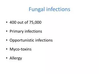
FUNGAL INFECTIONS • -Dermatophytoses • -Superficial infections of the skin. • -Common types are: • -tinea capitis • -tinea corporis • -tinea cruris • -tinea pedis
TINEA CORPORIS • -Ringworm of the body. • -Body parts that have little or no hair.
TINEA CRURIS • -Jock itch. • -Found in the groin area.
-Most common of all fungal infections. -Athlete’s foot. -Between the toes of people whose feet perspire heavily. -Contaminated swimming pools and public bathroom facilities TINEA PEDIS
SIGNS AND SYMPTOMS TINEA CAPITIS: • -erythematous. • -round lesion with pustules around the • edges • -temporary alopecia • -infected hairs will turn blue-green under a • Wood’s light.