Download
1 / 49
510 likes | 609 Vues
Explore the structure of liver and pancreatic tissues, including hepatic lobules and pancreatic islets. Learn about the exocrine and endocrine functions of the pancreas and the unique features of liver cells.

E N D
Liver and Pancreas Department of Histology and Embryology Medical college in Three Gorges University
Central vein hexagonal area
Basic Histology • In sections through the liver, the substance of the organ appears to be made up of hexagonal areas that constitute the hepatic lobules.
Hepatic lobules: (Central vein ,LM) (1)hepatic cords or plates (2)sinusoid (3) bile canaliculi In the section stained with H-E : only hepatic cords and sinusoids can be seen.
In transverse sections each lobule appears to be made up of cords of liver cells that are separated by sinusoids. However, the cells are really arranged in the form of plates (one cell thick) that branch and anastomose with one another to form a network. Spaces within the network are occupied by sinusoids.
The exocrine secretion of the liver cells is called bile. Bile is poured out from liver cells into very delicate bile canaliculi that are present in intimate relationship to the cells. From the canaliculi bile drains into progressively larger ducts which end in the bile duct.
portal area • Along the periphery of each lobule there are angular intervals filled by connective tissue, These intervals are called portal area.
Pepatic lobules 1.Hepatic cords: arranged one or two hepatic cell lines which are surrounding the central vein. LM: Each hepatocyte is a large cell with a round central nucleus, with prominent nucleoli. The cytoplasm is prominent. EM: abundant organelles
2. The sinusoids:capillary which lie between hepatic cords 1)lined by an endothelium in which there are numerous pores (fenestrate). 2)A basement membrane is not seen. good permeability. There are hepatic macrophages (Kupffer cells) in the sinusoids.
3. Perisinusoidal space: The surface of the liver cell is separated from the endothelial lining of the sinusoid by a narrow perisinusoidal space (of Disse). Microvilli, present on the liver cells, extend into this space. Ito cells: fat-storing cells are also seen in the space.
4.Bile canaliculi: They are merely spaces present between plasma membranes of adjacent liver cells. These canaliculi have no walls of their own. LM: nitrate siliver: black, network
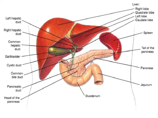
Portal area angular intervals filled by connective tissue. It contains: (a) a branch of the portal vein; (b) a branch of the hepatic artery (c) an interlobular bile duct These three structures collectively form a portal triad.
branch of the portal vein an interlobular bile duct branch of the hepatic artery
branch of the hepatic artery an interlobular bile duct branch of the portal vein
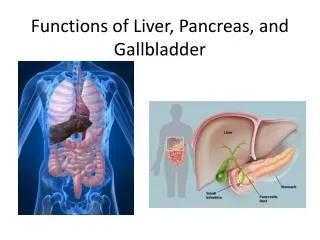
THE PANCREAS: The pancreas is a gland that is partly exocrine, and partly endocrine, the main bulk of the gland being constituted by its exocrine part.The exocrine pancreas secretes enzymes that play a very important role in the digestion of carbohydrates, proteins and fats.
Exocrine Part It is in the form of a serous, compound tubulo-alveolar gland. Its general structure is very similar to that of the parotid gland.
pancreatic islets Excroine part
serous acini centroacinar cell Secreting unit Exocrine Part Intercalated duct Intralobular duct Interlobular duct Pancreas duct pancreas Duct: Islet of Langerhans: A-cell,B-cell,D-cell, Endocrine Part:
Exocrine Part : acini Their lumen is small. The lining cells appear triangular in section, and have spherical nuclei located basally. In sections stained with haematoxylin and eosin the cytoplasm is highly basophilic (blue) specially in the basal part.
These ducts are invaginated deeply into the secretory elements.
Exocrine Part: Duct From the intercalated ducts the secretions pass into larger, interlobular ducts. They finally pass into the duodenum through the main pancreatic duct and the accessory pancreatic duct.
Centroacinar cell intercalated ducts
The Endocrine Part: is in the form of numerous rounded collections of cells that are embedded within the exocrine part. These collections of cells are called the pancreatic islets, or the islets of Langerhans The human pancreas has about one million islets. They are most numerous in the tail of the pancreas.
The islets are very richly supplied with blood through a dense capillary plexus. The intervals between the capillaries are occupied by cells arranged in groups or as cords.
(a) A-cells(or The alpha cells ) secrete the hormone glucagon. They form about 20% of the islet cells. (b) B-cells(or The beta cells ) secrete the hormone insulin. About 70% of the cells are of this type. (c) D-cells(or The delta cells ) probably produce the hormones gastrin and somatostatin. Somatostatin inhibits the secretion of glucagon by alpha cells, and (to a lesser extent) that of insulin by beta cells.
In islets of the human pancreas the alpha cells tend to be arranged towards the periphery (or cortex) of the islets. In contrast the beta cells tend to lie near the center (or medulla) of the islet. Delta cells are also peripherally placed. PP cell