GI Bleeding Scan
GI Bleeding Scan. รศ.พญ. มลฤดี เอกมหาชัย หน่วยเวชศาสตร์นิวเคลียร์ ภาควิชารังสีวิทยา คณะแพทยศาสตร์ มหาวิทยาลัยเชียงใหม่. GI Bleeding. 1. Upper GI bleeding 2. Lower GI bleeding. Diagnostic Procedures. 1. Endoscopic examination 2. Selective angiography 3. GI bleeding scan.


GI Bleeding Scan
E N D
Presentation Transcript
GI Bleeding Scan รศ.พญ. มลฤดี เอกมหาชัย หน่วยเวชศาสตร์นิวเคลียร์ ภาควิชารังสีวิทยา คณะแพทยศาสตร์ มหาวิทยาลัยเชียงใหม่
GI Bleeding 1. Upper GI bleeding 2. Lower GI bleeding
Diagnostic Procedures 1. Endoscopic examination 2. Selective angiography 3. GI bleeding scan
Acute Lower GI Bleeding 1. Small bowel bleeding 2. Large bowel bleeding
Causes of Small Bowel Bleeding • Regional enteritis • Intestinal varices • Lymphoma • Heriditary telangiectasia • Meckel’s diverticulum etc.
Causes of Large Bowel Bleeding • Diverticulosis • Inflammatory diseases • Arteriovenous malformation • Angiodysplasia • CA colon, Polyps etc.
Acute Lower GI Bleeding 1. Rigid endoscopy (Proctosigmoidoscopy) 2. Selective angiography 3. GI bleeding scan
Algorithm for the Dx & Rx of Acute Lower GI Hemorrhage Acute Lower GI Bleeding Proctosigmoidoscopy Rectal fissure, Bleeding site not found Hemorrhoids, Polyps or CA etc. GI bleeding scan Surgery or Therapeutic angiography
Algorithm for the Dx & Rx of Acute Lower GI Hemorrhage GI bleeding scan Positive scan Negative scan Angiography , Medical treatment Transcatheter Rx Elective colonoscopy Persistent bleedingBarium studies, CT etc Surgery
GI Bleeding Scan 1. Tc99m-Sulfur Colloids 2. Tc99m-labeled Red Blood Cells
Tc99m-Sulfur Colloid Scan • Dose 10 mCi IV • Circulation half-time 2.5-3.5 min • Active bleeding within 15-30 min • Minimal bleeding rate 0.05-0.1 ml/min
Mechanism • To demonstrate an acute GI bleed • Extravasation of activity (increased uptake)
Negative Tc99m- Sulfur Colloid Scan • Normal uptake in - Liver - Spleen & - Bone marrow
Positive Tc99m- Sulfur Colloid Scan • Normal uptake in liver, spleen & bone marrow • Extravasated activity at bleeding site(s)
Tc99m-labeled Red Blood Cell Scan • Dose 20 mCi IV • Active or intermittent bleeding • Minimal bleeding rate 0.35 ml/min
Negative Tc99m-RBC Scan • High uptake in - Blood pool organs & - Great vessels of abdomen
Positive Tc99m-RBC Scan • Normal uptake in blood pool organs & great vessels • Extravasated activity at bleeding site(s)
Scintigraphic Criterias for Dx of GI Bleeding 1. Area(s) of extravasated activity (increased uptake) - Appearsand - Conforms to bowel anatomy
Scintigraphic Criterias for Dx of GI Bleeding • 2. Area(s) of extravasated activity - Persistence or - Increased in intensitywith time
Scintigraphic Criterias for Dx of GI Bleeding 3. Area(s) of extravasated activity can change in - Size - Configuration & - Location with time.
Scintigraphic Criterias for Dx of GI Bleeding 4. Area of extravasated activity does not initially present, but appears in later images.(intermittent bleeding)
Advantages of GI Bleeding Scan • Noninvasive • More sensitive • Low cost • Low radiation exposure • Active or intermittent GI bleeding • Venous or arterial bleeding
Dose 20 mCi IV More complicated Noninvasive Low radiation exposure Lower target/nontarget ratio Active or intermittent bleeding Bleeding rate >0.35 ml/min Venous or arterial bleeding Dose 10 mCi IV Simple Noninvasive Low radiation exposure Higher target/nontarget ratio Active bleeding Bleeding rate > 0.1 ml/min Venous or arterial bleeding Tc99m-RBCTc99m-S.Colloid
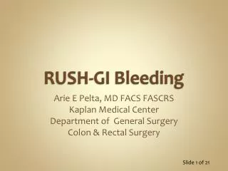
10 min 20 min
Ascending Colon Bleeding 10 min 20 min
5 min 30 min
Ascending Colon Bleeding 5 min 30 min
Negative Tc-99m RBC Scan 5 min 15 min 30 min 1 hour
Blood pool image Dynamic study 2sec/Frame
1 hr 2 hrs 3 hrs 4 hrs
Small Bowel Bleeding 1 hr 2 hrs 3 hrs 4 hrs
45 min 1 hour 2 hrs 3 hrs
Small Bowel Bleeding 45 min 1 hour 2 hrs 3 hrs