Lower GI Bleeding
Lower GI Bleeding. BY Dr. Waleed M. Abdel Maksoud MBBCh , MS, MD, MRCS (England). Definitions :.

Lower GI Bleeding
E N D
Presentation Transcript
Lower GI Bleeding BY Dr. Waleed M. Abdel Maksoud MBBCh, MS, MD, MRCS (England)
Definitions: • Melenais passage of digested blood (mainly mixed with bile and intestinal secretions), which is characteristically described as black tarry soft sticky offensive stools. It may cause a red halo around the soft fecal mass if mixed with water or urine.
Definitions: • Bleeding per rectum or hematochezia is passage of altered maroon colored or reddish dark brown colored offensive fluidy stool motions with possible clots. The nature of blood is evident to the patient.
Definitions: • Fresh bleeding per rectum is passage of fresh blood during passing a stool motion, and it takes several forms as splashing, drippling, streaking of stools or as terminal drops. Sometimes it stains the underwear after defecation. May be associated with mucus and /or pus
Lower GI Bleeding The ligament of Treitz is a musculofibrous band that extends from the upper aspect of the ascending part of the duodenum to the right crus of the diaphragm and tissue around the celiac artery. • Lower gastrointestinal bleeding is defined as abnormal hemorrhage into the lumen of the bowel from a source distal to the ligament of Treitz.
Common causes of massive lower gastrointestinal bleeding by age
Evaluation: • Lower GIT bleeding is considered massive and serious if: • 3-5 units of blood are needed over 24 hours to maintain stability. • if hematocrit is less than 30%. • if orthostatic changes in blood pressure occur.
Evaluation: • Evaluation is difficult for the following reasons: • Bleeding can originate from any area of the GIT. • Bleeding is often intermittent and source may be difficult to clarify if it is not actively bleeding. • This is a condition in which emergency surgery with significant morbidity and mortality may be required before specific diagnosis or even specific site of bleeding is identified. • Even radical types of surgery cannot prevent rebleeding. • There is no universal applicable sequence of investigations or treatments.
The basic components of management are • initial hemodynamic stabilization, • localization of the bleeding site, • site-specific therapeutic intervention
History • We should assess the chronicityof bleeding and medication use, particularly regarding: • anti coagulants such as • warfarin • low molecular weight heparin. • inhibitors of platelet aggregation such as • NSAIDs. • Clopidrogrel, this can associated with mesenteric ischemia. • Use of digitalis should be documented because this can associated with mesentricischemia.
Evaluation of the actively bleeding patient (1) Resuscitation: • with crystalloid solutions, preparing for blood transfusion. • diagnostic nasogastric intubation are important step to exclude Upper GI causes. The presence of bile in the aspirate is the only sure sign that there is no active upper GIT source of bleeding. (Upper endoscopy can diagnose more accurately the upper GIT source) • It should be recognized that about 85% of bleeding from lower GI sources stops spontaneously, however, the presence of large amounts of blood in the colon can give the impression of a continuous bleeding process. That is why in monitoring the bleeding, one should depend more on the hemodynamic figures rather than frequent passage of bloody motions.
Evaluation of the actively bleeding patient (2) Per rectal examination, anoscopy, and possibly rigid sigmoidoscopy: • are imperative to rule out palpable neoplasms and other anal and rectal conditions. • It is important, even if detection of a lesion is not possible due to bad preparation, to ascertain a healthy lower 15 or 20 cm for a possible use of subtotal colectomy. • Treatment of a simple visible cause like piles can be done at the same setting by injection or Baron band ligation.
Evaluation of the actively bleeding patient (3)Colonoscopy: • It offers the chance for proper diagnosis and sometimes-non-surgical stoppage of bleeding. • Mechanical preparation can be done and sometimes because the blood is very good laxative to the gut it is unnecessary. • It should be done as soon as the patient is stabilized and better to be done under intravenous general anesthesia.
Evaluation of the actively bleeding patient (3)Colonoscopy: • Diverticulosis, inflammatory bowel disease, polyps and neoplasms are confidently diagnosed, however arteriovenous malformations are sometimes difficult to diagnose, but if seen can be treated with injection of diluted adrenaline or photocoagulated by laser therapy. • The average detected lesions is in the range of 75%, about 85% of these are in the left colon, 10% in the right one. • Colonoscopy can also diagnose the source of bleeding as small bowel if the cecum is reached and more fresh blood is coming out of the ileocecal valve. This will indicate small bowel site, which is most propably ulcers, IBD, benign or malignant small bowel neoplasms (leiomyoma, leiomyosarcoma or lymphoma) Meckel’sdiverticulum, or arteriovenous malformation are also other possibilities.
Malignancy Colon Carcinoma
Video Capsule Endoscopy • Capsule endoscopy uses a small capsule with a video camera that is swallowed and acquires video images as it passes through the GI tract. • This modality permits visualization of the entire GI tract, but offers no interventional capability. • It is also very time consuming because someone has to watch the video to identify the bleeding source, and then a means to deal with the pathology has to be developed.
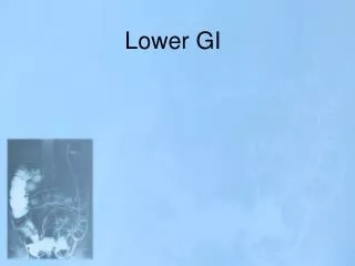
Evaluation of the actively bleeding patient (4) Arteriography: • By selective catheterization of all mesenteric vessels, and technetium labeled RBC scanning are only of use if active bleeding is present with a high rate (2ml/minute or higher), which is not usually the case on admission of the patient. • Selective vasopressin infusion or embolization with thrombin or gel foam can help to stop bleeding.
Angiographic study documents extravasation of contrast into small bowel. Intraoperative examination of bowel is aided by injection of methlyene blue dye, which facilitates localization of bleeding site and thereby helps direct surgical resection.
Evaluation of the actively bleeding patient (5) Surgical intervention: • Indications: • Failure of all preveuos measures to stop bleeding. • When the bleeding is massive (blood loss more than 2.5 litres over 48 hrs). • In these situation, surgical intervention will have a lower morbidity than continued conservative management.
Evaluation of the actively bleeding patient (5) Surgical intervention: • Procedure: • If the source of bleeding could be localized preoperatively or intra-operatively, segmental resection would be performed. • If the source of bleeding could not be localized preoperatively or intra-operatively: • Intraoperativeenteroscopy is reserved for patients who have transfusion-dependent obscure-overt bleeding in whom an exhaustive search has failed to identify a bleeding source. • Segmentation of the colon is done by means of non crushing clamps and observation of the segment that will collect blood. • If all measures fail to localize the source of bleeding, sub-total colectomy may be indicated.