Download
1 / 12
240 likes | 1.22k Vues
Approach to a Patient with Lower GI Bleeding. Antonio. Aramburo. Arcilla. Argana. Patient. L. Q. 78 y/o Female Chief Complaint: Hematochezia. Salient Features:. Chief Complaint: Hematochezia 6 hours PTA- ½ teaspoon of blood after defecation 4 hours PTA- 1 tablespoon of blood

E N D
Approach to a Patient with Lower GI Bleeding Antonio. Aramburo. Arcilla. Argana
Patient • L. Q. • 78 y/o • Female • Chief Complaint: Hematochezia
Salient Features: • Chief Complaint: Hematochezia • 6 hours PTA- ½ teaspoon of blood after defecation • 4 hours PTA- 1 tablespoon of blood • 30 mins PTA- 2 cupfuls of fresh blood -Dizzy, cold clammy perspiration
Approach to the Patient: Lower Gastrointestinal Bleeding • Measure the heart rate and blood pressure
Approach to the Patient: Lower Gastrointestinal Bleeding • Differentiation of upper from lower GIB • Hematemesis- indicates upper GI source of bleeding • Hematochezia- usually represents lower GI source of bleeding
Approach to the Patient: Lower Gastrointestinal BleedingDiagnostic Evaluation of the Patient with Lower GIB Upper endoscopy – to rule out an upper GI source before evaluation of lower GI tract -Patients with hematochezia and hemodynamic instability
Diagnostic Evaluation of the Patient with Lower GIB • Sigmoidoscopy • for patients <40 years old with minor bleeding • for detection of obvious, low-lying lesions • risk of bleeding, area of bleeding is usually not possible to identify
Diagnostic Evaluation of the Patient with Lower GIB • Colonoscopy- procedure of choice
Diagnostic Evaluation of the Patient with Lower GIB • Tc-labeled red cell scan -allows repeated imaging for up to 24 hours - may identify the general location of bleeding
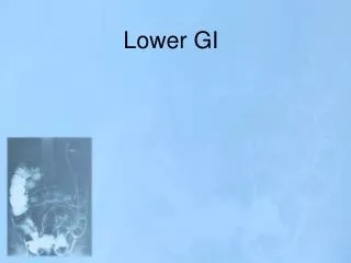
Diagnostic Evaluation of the Patient with Lower GIB • Angiography - can detect the site of bleeding - permits treatment with intraarterial infusion of vasopressin or embolization - may identify lesions with abnormal vasculature, such as tumors or vascular ectasias
Figure 1.1 Suggested algorithm for patients with acute lower gastrointestinal bleeding
Differential Diagnosis • Common causes of LGIB • Diverticula • Vascular ectasia (Angiodysplasia) • Neoplasms (Adenocarcinoma)