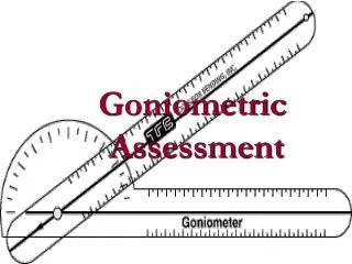
Goniometric Assessment
Goniometric Assessment. Joints . NASM only chose a select number of joints to be measured Foot Dorisflexion Hip Flexion (Bent knee and 90/90 position) Internal Rotation External Rotation Extension Abduction Shoulder Flexion External Rotation Internal Rotation

Goniometric Assessment
E N D
Presentation Transcript
Joints • NASM only chose a select number of joints to be measured • Foot • Dorisflexion • Hip • Flexion (Bent knee and 90/90 position) • Internal Rotation • External Rotation • Extension • Abduction • Shoulder • Flexion • External Rotation • Internal Rotation • Measurements were selected because of their overall importance to optimum human movement as well as their ability to correlate to the overhead squat and single movement assessment.
The Foot • Joint motion being assessed • Dorsiflexion of talocrural joint • Muscles being assessed • Gastrocnemius and soleus • Posterior tibialis, peroneus longus, flexor hallicus longus, and flexor digitorum longus. • Antagonists potentially underactive if ROM is limited • Anterior tibialis • Extensor digitorum longus, extensor digitorum brevis, extensor hallicus longus and peroneus tertius. • Normal Value- 20o • Client Positioning • Supine with Knee extended • Ankle is subtalar neutral
Placement of Goniometer • Axis (A)- Directly below the lateral mallelous near the base • Stationary Arm (SA) – Lateral aspect of fibula • Movement Arm- (MA) Midline of 5th metatarsal. • Pressure • Hold planter surface of foot right below MTP joints • Client/Patient actively DF while you are passively assisting the glide of motion • Compensation during Goniometer Assessment • Everson of the ankle • Flexing of the knee • Over Head Squat/ Single Leg Squat • Foot compensations ( feet going outward Flattening and/or heels rising) • Excessive forward leaning • A lack of DF in the ankle has been know to lead to knee injuries.
Hip Flexion • Joint motion being assessed • Extension of the tibiofemoral joint • Flexion of iliofemoral joint • Muscles being assessed • Hamstrings, Gastrocnemius, neural tissue (sciatic nerve) • Antagonists potentially underactive if ROM is limited • Hip flexor complex • Quadriceps complex • Normal Value- 20o • Client Positioning • Supine with Hip flexed and knee flexed to 90o • Hip is in neutral (0o rotation, abduction and adduction)
Placement of Goniometer • Axis (A)- lateral joint line of the tibiofemoral joint • Stationary Arm (SA) – Lateral midline of femur • Movement Arm (MA)- lateral midline of fibula • Pressure • Hold lower leg and thigh of client • Passively extend the knee until first compensations • Compensation during Goniometer Assessment • Posterior tilting of the pelvis • Hip extension • Over Head Squat/ Single Leg Squat • Feet turned out (External rotated) • Feet flattening • Knee moving inward or outward • Low back rounding
Hip Flexion (Bent Knee) • Joint motion being assessed • Flexion of iliofemoral joint • Muscles being assessed • Gluteus maximus, adductor magnus, upper portion of hamstrings • Psoas, rectus femoris, hip capsule. • Antagonists potentially underactive if ROM is limited • Hip flexor complex • Hip extensor complex (gluteus maximus) • Normal Value- 120o • Client Positioning • Supine with knee flexed • Hip is in neutral (0o rotation, abduction and adduction)
Placement of Goniometer • Axis (A)- Great trochanter • Stationary Arm (SA) – Lateral midline of pelvis • Movement Arm (MA)- lateral midline of femur • Pressure • Hold clients knee • Passively flex the hip until first compensation. • Compensation during Goniometer Assessment • Posterior tilting of the pelvis • Adbuction of the femur • Over Head Squat/ Single Leg Squat • Rounding of the lower back
Hip (Internal Rotation) • Joint motion being assessed • Internal rotation of iliofemoral joint • Muscles being assessed • Piriformis and hip external rotators and adductor magnus, ischiofemoral ligaments • Gluteus medius, gluteus maximus • Antagonists potentially underactive if ROM is limited • Adductor magnus, TFL, gluteus minimus, glutues medius, adductor longus, adductor brevis, pectineus, gracilis, medial hamstrings. • Normal Value- 45o • Client Positioning • Supine with Hip flexed and knee flexed to 90o • 0o of abduction and adduction
Placement of Goniometer • Axis (A)- Anterior aspect of patella • Stationary Arm (SA) – parallel to imaginary line down the center of the body • Movement Arm (MA)- Anterior midline of the lower leg (referencing the tibial tuberosity). • Pressure • Hold lower leg and thigh of client • Passively rotate the femur internally until first compensation • Compensation during Goniometer Assessment • Hip hike ( lateral flexion of spine) on side of measurement • Over Head Squat/ Single Leg Squat • Knee moving inward or outward • Asymmetrical weight shift
The Hip (External Rotation) • Joint motion being assessed • External rotation of iliofemoral joint • Muscles being assessed • Adductor magnus, iliofemoral ligament, and pubofemoral ligament • TFL, gluteus minimus, and gluteus medius • Antagonists potentially underactive if ROM is limited • Piriformis and hip external rotators and adductor magnus • Gluteus medius and gluteus maximus. • Normal Value- 45o • Client Positioning • Supine with hip and knee flexed to 90o
\ • Placement of Goniometer • Axis (A)- Anterior aspect of patella • Stationary Arm (SA) – parallel to imaginary line down the center of the body • Movement Arm (MA)- Anterior midline of the lower leg (referencing the tibial tuberosity). • Pressure • Hold lower leg and thigh of client • Passively rotate the femur externally until first compensation • Compensation during Goniometer Assessment • Motion of ASIS • Over Head Squat/ Single Leg Squat • Knee moving inward or outward • Asymmetrical weight shift
Hip (Extension) • Joint motion being assessed • Extension of iliofemoral joint • Muscles being assessed • Psoas, iliacus, rectus femoris, tensor fascia latae and sartorius • Adductor complex and anterior hip capsule • Antagonists potentially underactive if ROM is limited • Gluteus maximus, glutues medius • Hamstring complex, adductor magnus • Normal Value- 0-10o • Client Positioning • Supine with opposite hip flexed • Knee of testing leg should be flexed to ~ 90o
Placement of Goniometer • Axis (A)- Greater Trochanter • Stationary Arm (SA) – lateral midline of the trunk • Movement Arm (MA)- Lateral midline of the femur • Pressure • Hold thigh of client • Passively allow the hip to extend until first compensation. • Compensation during Goniometer Assessment • Anterior tilting • Low back arching • Over Head Squat/ Single Leg Squat • Arching of the lower back • Excessive forward lean
Hip (Abduction) • Joint motion being assessed • Abduction of iliofemoral joint • Muscles being assessed • Adductor complex, pubofemoral ligament, iliofemoral ligament, medial hip capsule • Medial Hamstrings • Antagonists potentially underactive if ROM is limited • Gluteus medius, Gluteus minimus, TFL, Satorius • Bicep Femoris • Normal Value- 40o • Client Positioning • Supine with knee extend • Hip is neutral
Placement of Goniometer • Axis (A)- ASIS • Stationary Arm (SA) – Imaginary line b/w ASIS’s • Movement Arm (MA)- Anterior midline of femur • Pressure • Holding Clients lower leg • Passively abduct the leg until first compensation • Compensation during Goniometer Assessment • Motion of opposite ASIS • Hip Hike on side of movement • Over Head Squat/ Single Leg Squat • Knees moving inward • Asymmetrical weight shift
Shoulder (Flexion) • Joint motion being assessed • Flexion of Shoulder complex • Muscles being assessed • Latissimus dorsi, teres major, teres minor, infraspinatus, subscapularis, pectoralis major, triceps • Antagonists potentially underactive if ROM is limited • Anterior deltoid, pectoralis major, middle deltoid • Lower and middle trapezius, rhomboids. • Normal Value- 160o • Client Positioning • Supine with should neutral • Knee’s in hook-lying position • Arm in external rotation
Placement of Goniometer • Axis (A)- Distal to the acromion process • Stationary Arm (SA) – mid-axillary line of upper thorax • Movement Arm (MA)- Lateral epicondyle of the humerus • Pressure • Hold arm in external rotation • Place thumb on the lateral border of the scapula and passively flex the shoulder until excessive scapular movement is felt or resistance is felt. • Over Head Squat/ Single Leg Squat • Arching of the lower back • Arms falling forward
Shoulder (External Rotation) • Joint motion being assessed • External rotation of glenohumeral joint • Muscles being assessed • Subscapularis, latissimus dorsi, teres major, pectoralis major, anterior deltoid and anterior glenohumeral joint capsule. • Antagonists potentially underactive if ROM is limited • Infraspinatus, teres minor, posterior glenohumeral joint capsule • Normal Value- 90o • Client Positioning • Supine with humerus abducted to 90o • Elbow flexed to 90o • Towel is placed under humerus
Placement of Goniometer • Axis (A)- Olecranon process • Stationary Arm (SA) – Perpendicular to the arm • Movement Arm (MA)- Ulnar styloid • Pressure • Hold arm in external rotation till first resistance • Compensation during Goniometer Assessment • Upward migration of the humeral head into the hand over the anterior shoulder. • Over Head Squat/ Single Leg Squat • Arms falling forward
Shoulder (Internal Rotation) • Joint motion being assessed • Internal rotation of glenohumeral joint • Muscles being assessed • Infraspinatus, teres minor, posterior glenohumeral joint capsule • Antagonists potentially underactive if ROM is limited • Subscapularis, latissimus dorsi, teres major, pectoralis major, anterior deltoid. • Normal Value- 70o • Client Positioning • Supine with humerus abducted to 90o • Elbow flexed to 90o • Towel is placed under humerus
Placement of Goniometer • Axis (A)- Olecranon process of elbow • Stationary Arm (SA) – Perpendicular to the floor • Movement Arm (MA)- Ulnar styloid and olecranon process • Pressure • Hold arm in internal rotation until first resistance. • Compensation during Goniometer Assessment • Upward migration of the humeral head into the hand over the anterior shoulder. • Over Head Squat/ Single Leg Squat • Arms falling forward
Reference • National Academy of Sports Medicine. Goniometric assessments. California, 2005 (1-38).