Chapter 4 Cartilage and Bone
Chapter 4 Cartilage and Bone. 1. Cartilage: organ=Cartilage tissue+perichondrium . 1) structure of cartilage tissue ---cell: chondrocyte ---cartilage matrix. ① chondrocyte: ---Structure: LM embedded in cartilage lacuna peripheral cells:

Chapter 4 Cartilage and Bone
E N D
Presentation Transcript
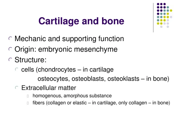
1. Cartilage: organ=Cartilage tissue+perichondrium
1) structure of cartilage tissue ---cell: chondrocyte ---cartilage matrix
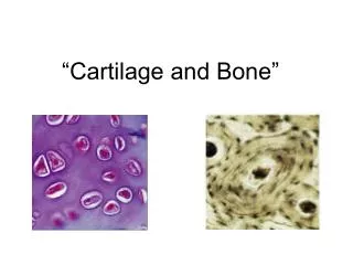
① chondrocyte: ---Structure: LM • embedded in cartilage lacuna • peripheral cells: --small and immature --single and flattened • central cell: --large and mature, --round and in group of 2-8 cells --small and round nucleus --basophilic cytoplasm --EM: rich in RER and Golgi complex
*isogenous group: several cells locates in one lacuna, which are derived from a single(same) parent cell
② Cartilage matrix ---ground substance: • proteoglycan: --same to loose CT --there are more chondroitin sulfate distributed at the periphery of cartilage lacuna---called as cartilage capsule(basophilic) • chondronectin • water ---fiber: type and number of fiber depends on the type of cartilage
2) Classification: according to the fiber • Hyaline cartilage: • less collagenous fibril←type II collagen • articular surface, rib cartilage, trachea and bronchi
Fibrous cartilage: • large amount of collagenous fiber bundles • cells are small and less • intervertebral disc, symphysis pubis
Elastic cartilage: • large amount of elastic fiber • external ear, epiglottis
3) perichondrium two layers: ---out layer: contain more fiber-protection ---inner layer: more cells-osteoprogenitor cell(fusiform in shape)
4) growth of cartilage ---interstitial growth: • inner chondrocyte proliferation→ produce fiber and matrix. • immature cartilage ---appositional growth: • osteoprogenitor cell→cartilage cell (chondrocyte) → produce fiber and matrix. • growing and mature cartilage
2.Bone ---consists of bone tissue, periosteum and endosteum, bone marrow
1)Bone tissue ① Cells:
a. osteoprogenitor cell: stem cell ---structure: • fusiform, small • ovoid nucleus • slight basophilic cytoplasm • exist in periosteum and endosteum ---function: differentiated into osteoblast and chondrocyte
b. osteoblast: ---structure: LM: • single layer of cuboidal or low columnar cell • round nucleus • basophilic cytoplasm • located on the surface of bone tissue
EM: • fine processes • rich in RER, Golgi complex
---function: ⅰ.synthesize bone collagen fiber and ground substance-osteoid ⅱ.release matrix vesicle: • 0.1um in diameter • membrane-coated • ALPase(Alkaline phosphatase), ATPase and pyrophosphatase and phosphoester (phospholipid) • calcium, crystal of bone salt and calbindin • function: promote calcification
c.osteocyte ---structure: • flattened cell with multiple long thin processes • located in bone lacuna and bone canaliculus • basophilic cytoplasm • adjacent cells connect in bone canaliculus by gap junctions ---function: • Maintain bone matrix • regulate the balance of calcium and phosphonium
d. osteoclast ---structure: LM: • multinuclear large cell, 30-100um • 6-50 nuclei • acidophilic cytoplasm • located at peripheral part of bone
EM: • ruffled border-processes • light zone: --under the ruffled border --microfilament • primary lysosome, pinosome and secondary lysosome • RER, mito. and Golgi ---function: dissolve and absorb bone matrix
②Bone matrix ---organic matter: • bone collagen fiber -collagenous fiber (type I collagen) • ground substance: glycosaminoglycan
glycoproteins: • osteocalcin: involve in calcification of bone and regulate absorption of bone • osteonectin: related to adherence between cell and bone matrix, regulate calcification of bone • osteopontin
---inorganic matter: bone salts Hydroxyapatite crystal: • Ca10(PO4)6(OH)2 • pin-shaped • 10-20 nm • longitudinal arranged *bone lamella: bone matrix arranged in layers at different direction
2) Architacture of long bone Long bone is an organ, made up of bone tissue(shaft and epiphyses), periosteum and endosteum, bone marrow
① shaft: consists of compact bone a.circumferential lamella: /outer concentrically-arranged /inner around inner surface of bone
b.Haversian system (osteon): /cylindric structure, 3-5mm /central canal: N, BV, CT /Haversian lamella: 4-20 layers
c.interstitial lamella: /irregular lamella /remnant of Haversian or circumferential lamella *perforating canal: /transverse canal /connect with Haversian canal
② epiphyses: composed of spongy bone ---trabeculae: • formed by parallelly-arranged lamella • form a spongy-liked network ---Bone marrow: hemopoietic tissue
③ periosteum and endosteum: CT membrane ---periosteum: DCT • outer layer:more fiber bundles form perforating fiber • inner layer: rich in BV, N and osteoprogenitor cells ---endosteum: thin, a layer of osteoprogenitor cell and CT ---function: provide nutrition and osteoblast for bone growth and repairing
①basal processes ---formation: osteoprogenitor cell→ osteoblast → osteoid ↓ ↓calcification osteocyte + bone matrix bone tissue ---absorption: osteoclast →dissolve bone tissue→reconstruction
②basal manner a. intramembranous ossification: ---CT membrane →osteoprogenitor cell → osteoblast→ossification center→bone trabeculae →thicker and longer ---flattened bone and irregular bone formed in these manner
b. endochondral ossification: e.g. long bone ⅰ.formation of cartilage model • Mesenchymal cell→osteoprogenitor cell →chondroblast→chondrocyte→cartilage model( consists of hyaline cartilage and perichondrium) ⅱ.formation of bone collar • osteoprogenitor cell (perichondrium) → osteoblast →bone tissue * These bone tissue surround the central segment of cartilage model as collar-shaped, so called bone collar
ⅲ.formation of primary ossification center and bone marrow cavity • chondrocytes of model center stop differentiation, enlarge in size, calcification, dead →CT, BV in periosteum enter degenerating zone→osteoblast, osteoclast, osteoprogenitor cell and mesenchymal cell enter at same time→ossification→primary ossification center
primary bone marrow cavity(space between trabeculae) →bone marrow cavity
ⅳ.Formation of secondary ossification center and epiphyses • secondary ossification center appears at the two end of long bone(epiphyses) • epiphyseal plate: cartilage layer between epiphysis and bone shaft, growing zone
③Further growth of bone ---Become longer: • by growth of epiphyseal plate • from epiphyses to shaft, four zones can be seen: i.reserve cartilage zone: cell is small, round and basophilic ii.proliferating cartilage zone: cell is flattened, isogenous group cell arrange in single line
iii. calcified cartilage zone: cell become large, mature, round and degenerated, strong basophilic iv.ossification zone: ---become thicker: periosteum cell → osteoprogenitor cell→osteoblast