Download
1 / 80
830 likes | 1.14k Vues
Knee. Brandon Mines, MD Emory Sports Medicine Center. Anatomy Physical exam Special testing Injuries & how they relate to the above. Objectives. Don’t feel overwhelmed! Develop systematic examination Try not to skip regions of the knee Don’t forget joint above and below. Knee.

E N D
Knee Brandon Mines, MD Emory Sports Medicine Center
Anatomy Physical exam Special testing Injuries & how they relate to the above Objectives
Don’t feel overwhelmed! Develop systematic examination Try not to skip regions of the knee Don’t forget joint above and below Knee
Acute or chronic injury? Effusion present? Mechanism of injury? Aggravating/alleviating factors? Important areas to address
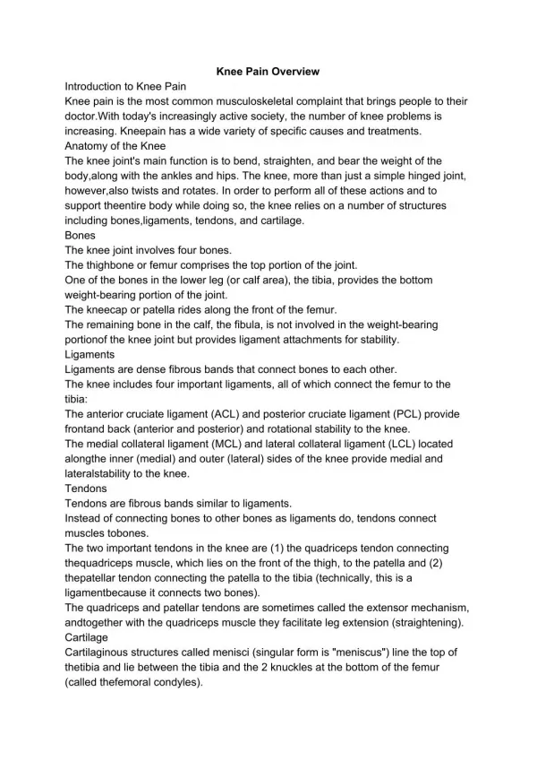
Knee overview • Knee pain ~ 33% of MSK problems seen in primary care clinics • Up to 55% of athletes complain of knee pain in a given year
As usual, a thorough, complete history is crucial • Pain • Onset rapid or insidious • Where is it located • How long has it been present • What is the severity & quality • Aggravating & alleviating factors • Bear weight immediately or not
History • Mechanical symptoms • Locking or catching • Popping (at injury and/or now) • Giving way
History • Effusion • Is there/was there one • Rapid (< 1 hour) • Delayed (24-36 hours)
History • Mechanism of Injury • Direct blow & location • Twisting, landing, cutting, decelerating • Planted foot • Unknown
Medical history Previous injury or hx of surgery Meds used to treat sxs Physical therapy Braces or other devices used Hx of gout, RA, DJD History
Physical Exam: Inspection • First, be sure to adequately visualize both knees • Inspection • Anatomic deformities • Gait abnormalities • How they disrobe
Physical Exam: Inspection • Observe both knees for erythema, swelling, bruising • Observe quad muscle carefully for possible atrophy
Physical Exam • Examine uninjured knee first to keep patient at ease • Save most ‘obnoxious’ maneuvers until the end
Physical Exam: Effusion • Milk knee to assess for effusion • Squeeze medial & lateral while milking
Physical Exam: Effusion • Ballotable patella
Physical Exam: Effusion • Compare this with ‘good’ knee • Intra-articular vs bursal Bursal Intra-articular
Patellofemoral assessment • Patella apprehension test
Patellofemoral assessment • Important to assess how the patella moves when the knee is flexed & extended Patellar tracking
Patellofemoral assessment • Patellar mobility • Palpate superior, inferior, medial & lateral patella facets
Patellofemoral assessment • Integrity of patellar & quad tendon • Compression test • Patellar inhibition
Range of Motion • Note angle of knee while supine • Passive ROM • Can you make the knee fully extend? • Is there full flexion? Is it limited by pain or mechanical cause?
Anterior knee palpation • Flex knee to 90° • Palpate medial & lateral joint lines • Palpate MCL & LCL • Palpate tibial tubercle Medial knee Lateral knee Tibial tubercle
Ligament testing • ACL testing • Lachman’s - 30° flexion • Anterior Drawer - 90° flexion Lachman’s Anterior drawer
Ligament testing • PCL testing • Posterior drawer test
Ligament testing • MCL testing • Valgus stress test • 0° & 30°
Ligament testing • LCL testing • Varus stress test • 0° & 30°
Meniscus • Meniscus testing • McMurray test
Patellofemoral pain syndrome • Retropatellar or peripatellar pain resulting from physical or biomechanical changes in the patellofemoral joint • Many forces interact to keep the patella aligned
Patellofemoral pain syndrome • Patella not only moves up and down, but rotates and tilts • Many points of contact between patella and femoral structures
Patellofemoral pain syndrome • Hx: • Vague anterior knee pain with insidious onset • Common cause of anterior knee pain in women • Tend to point to front of knee when asked to localize pain • Worse with certain activities, i.e. ascending or descending hills & stairs • Pain with prolonged sitting → theater sign • No meniscal or ligamentous sxs
Patellofemoral pain syndrome • PE: • Positive compression test • Patellar crepitus with ROM • Mild effusion possible • May see tenderness with patella facet palpation → medial, lateral, superior, inferior • Remainder of knee exam unremarkable
Patellofemoral pain syndrome • PE: • Check hamstring flexibility
Patellofemoral pain syndrome • PE: • Check for flat feet (pes planus) or high-arch feet (pes cavus) Pes Planus Pes Cavus
Patellofemoral pain syndrome • PE: • Check heel cord (achilles) flexibility • Check for a tight iliotibial band (ober’s test) Ober’s test Achilles stretch
Patellofemoral pain syndrome • Tx: • Physical therapy • Improve flexibility • Quad strengthening, especially VMO • Other modalities, i.e. soft tissue release, U/S • Patellar taping
Patellofemoral pain syndrome • Tx: • Relative rest/Modification of activities • Icing • NSAIDS • Patellar braces • Addressing foot problems with foot wear and orthotics • Surgery
Iliotibial band tendonitis • Excessive friction between iliotibial band (ITB) & lateral femoral condyle
Iliotibial band tendonitis • Common in runners and cyclists • Tight ITB, foot pronation, genu varum are risk factors
Iliotibial band tendonitis • Hx: • Pain at lateral knee • At first, sxs only after a certain period of activity • Progresses to pain immediately with activity
Iliotibial band tendonitis • PE: • Tender at lateral femoral epicondyle, ~3cm proximal to joint line • Soft tissue swelling & crepitus • No joint effusion
Iliotibial band tendonitis • PE: • Ober’s test • Noble’s test Noble’s test
Iliotibial band tendonitis • Tx: • Relative rest • Ice • NSAIDS • Stretching • Cortisone • Platelet-Rich Plasma
Iliotibial band tendonitis • Prognosis: • Improves with rest • Expect long recovery time • When to refer: • Intractable pain • Surgery = release
Anterior cruciate ligament (ACL) injury • Most are non-contact injury, 2° to deceleration forces or hyperextension • Planted foot & sharply rotating • If 2° to contact, may have associated injury (MCL, meniscus)