Anatomy and Physiology Course Syllabus Overview - Spokane Falls Community College
This syllabus provides essential details about the Anatomy and Physiology course at Spokane Falls Community College, taught by Jessica Radke-Snead. The schedule includes lectures and lab sessions on Mondays and Wednesdays, exam dates, and attendance expectations. Students are encouraged to actively participate, maintain academic integrity, and seek help when needed. The syllabus outlines grading criteria based on points from lectures, labs, and quizzes. Key topics include organ systems, cell types, and epithelial tissue, vital for the course curriculum.

Anatomy and Physiology Course Syllabus Overview - Spokane Falls Community College
E N D
Presentation Transcript
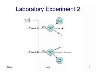
Laboratory 2 Jessica Radke-Snead, RD, MS Anatomy and Physiology 241 Spokane Falls Community College
Syllabus Review • Schedule: Mon/Wed 300-500pm • 4 Lecture and Lab Exams: Jan 16, Feb 6, Feb 27 and Mar 18 • No Lab on Jan 21 or Feb 18 • Attendance • Expectation is that you will attend class • Successful students attend class, are prepared for class and are active participants in the class • Missed information and missed opportunities affects test performance, as well as individual professional integrity • Un-announced quizzes may be given at any time • Absences • Lecture exams must be made up within one week • Lab exams cannot be made up—no exceptions • Find another lab time to sit for the exam if you can’t make yours
Syllabus Review • Grades • Earned on a basis of total points derived from both lecture and lab exams, and any appointed assignments or quizzes • A detailed grading scale will be posted online to track your progress • Personal honestly and integrity • The most important attributes of any professional • Cheating will not be tolerated. Period.
Syllabus Review • Student responsibilities • Behave in a professional manner • Pay attention to course calendar • Actively participate in course activities • Seek assistance from instructors when needed • Take control of your attitude, time and performance • Instructor responsibilities • Behave in a professional manner • Facilitate a positive learning environment and open-communication • Guide students in their quest to gain knowledge • Establish well-defined student goals • Share knowledge • Provide a course calendar with specific dates of events • Inform students of their performance and grades in a timely manner
Syllabus Review • Tips for success • Be organized and efficient • Start small and build your foundation • Begin with terms, connect terms to make a concept, connect concepts to understand the big picture • Study lecture and lab materials simultaneously—make connections, develop the “big picture” • Regularly and effectively study and review course materials • Isolate study time, eliminate distractions • Make a plan and see it through
Lab 1 Review: Organ Systems What organ systems are primarily involved in: Protection, support and movement? Internal communication and integration? Fluid transport? Defense? Input and output? Reproduction?
Organ systems: Answers Protection, support and movement: Integumentary, skeletal and muscular systems Internal communication and integration: Nervous and endocrine system Fluid transport: Circulatory and lymphatic system Defense: Immune system Input and output: Respiratory, urinary and digestive systems Reproduction: Reproductive system
Lab 1 Review: True or False? • A single sagittal section of the body can pass through one lung but not through both. • It is possible to see both eyes in one frontal section of the head. • The knee is both superior and proximal to the tarsal region. • The diaphragm is ventral to the lungs. • The esophagus is in the dorsal body cavity. • The liver is in the lateral abdominal region. • The heart is in the mediastinum. • Both kidneys could be shown in a single coronal section of the body. • The peritoneum lines the inside of the stomach and intestines. • The sigmoid colon is in the lower right quadrant of the abdomen.
True or False: Answers • A single sagittal section of the body can pass through one lung but not through both. TRUE • It is possible to see both eyes in one frontal section of the head. TRUE • The knee is both superior and proximal to the tarsal region. TRUE • The diaphragm is INFERIOR to the lungs. • The esophagus is in the VENTRAL body cavity. • The liver is in the HYPOCHONDRIAC region, which is superior to the lateral abdominal region. • The heart is in the mediastinum. TRUE • Both kidneys could be shown in a single coronal section of the body. TRUE—FRONTAL = CORONAL • The peritoneum lines the OUTSIDE of the stomach and intestines. • The sigmoid colon is in the lower LEFT quadrant of the abdomen.
Lab 2: Microscope, Cells and Epithelial Tissue • Common cell shapes • Squamous: thin, flat, angular contours (esophageal lining, skin) • Cuboidal: squared (liver) • Columnar: rectangular; markedly taller than wide (intestinal lining) • Spheroid/Ovoid: round/oval (fat and egg cells) • Discoid: doughnut-shaped (RBCs) • Stellate: multiple extensions, star-like shape (nerve cells) • Fusiform (spindle-shaped): thick center, tapered ends (smooth muscle cells) • Fibrous: thread-like shape (skeletal muscle cells)
Squamous Epithelium Description: • Single (simple) or multiple layers (stratified) of flat, scale-like cells • Spherical to stretched, centrally placed nucleus What are these adapted for? Diffusion and filtration Where do we find them? Endothelium (lines heart and BV) and mesothelium (lines thoracic and abdominopelvic cavities, covers organs within these cavities as part of serous membranes)
Stratified Squamous Epithelium Description: • Multiple cell layers with cells becoming increasingly flat toward surface • Spherical to stretched, centrally placed nucleus • Basal cells may be cuboidal to columnar What are these adapted for? • Resists abrasion (“sloughs off”) • Penetration by pathogenic organisms Where do we find them? Tongue, oral mucosa, esophagus, anal canal, vagina
Keratinized Stratified Squamous Epithelium Description: • Multiple cell layers with cells becoming increasingly flat toward surface • Basal cells may be cuboidal to columnar • Additional surface layer of keratin (protein resistant to friction that repels bacteria) and compact dead cells without nuclei What are these adapted for? Resists abrasion, retards water loss through skin and resists penetration by pathogenic organisms Where do we find them? Epidermis; Palms of hands and bottom of feet are especially heavily keratinized
Cuboidal Epithelium Description: • Typically single layer of square cells • Spherical, centrally placed nucleus • Pyramidal and arranged around a central space in glands • Brush border of microvilli in some kidney tubules • Ciliated in bronchioles of lung What are these adapted for? • Absorption and secretion • Production of protective mucous coat • Movement of respiratory mucus Where do we find them? Liver, thyroid, mammary, salivary and other glands, most kidney tubules and bronchioles
Columnar Epithelium Description: • Tall, narrow cells • Oval-shaped nuclei, vertically oriented, usually in the basal half of the cell • Often shows a brush border of microvilli—ciliated in some organs • May possess goblet cells What are these adapted for? • Absorption • Secretion of mucus and other products • Movement of egg and embryo in uterine tube Where do we find them? Inner lining of stomach, intestines, gallbladder, uterus and uterine tubes; some kidney tubules
Non-Ciliated Columnar Epithelium Description: • Contains a brush border of microvilli What are these adapted for? • Increases the surface area and rate of absorption • Goblet cells secrete mucus Where do we find them? Inner lining of stomach, and intestines
Pseudostratified Columnar Epithelium Description: • Appears multi-layered • Some cells do not reach the surface, but all cells reach the basement membrane • Nuclei at several levels in deeper half of epithelium • Often with goblet cells • Often ciliated What are these adapted for? • The cells that reach the surface either secrete mucus (goblet cells) or bear cilia that sweep away mucus and trapped foreign particles Where do we find them? Respiratory tract from nasal cavity to bronchi, portions of male urethra
Transitional Epithelium Description: • Resembles stratified squamous epithelium, but surface cells are rounded, not flattened and often bulge at surface • Typically 5-6 cells thick when relaxed and 2-3 cells thick when stretched • Cells may be flatter and thinner when epithelium is stretched (distended bladder) • Some cells have 2 nuclei What are these adapted for? • Stretches to allow filling or urinary tract Where do we find them? Urinary tract-portion of kidney, ureter, bladder, part of urethra, allantoic duct in umbilical cord
Cell Surface Area and Volume • Most human cells range from 10-15 micrometers in diameter • As a cell doubles in diameter, its volume increases 8-fold but its surface area only increases 4-fold. • Effect of cell growth • Diameter (D) is increased by a factor of 2 (D x 2) • Surface area is increased by a factor of 4 (D2) • Volume is increased by a factor of 8 (D3) A cell that is too large may have too little plasma membrane to serve the metabolic needs of its increased volume of cytoplasm, ie it cannot support itself
Mitosis: The Cell Cycle • Length of cell cycle varies greatly from one cell type to another • Stomach and skin cells divide rapidly—why? • Bone and cartilage cells divide slowly—why? • Skeletal muscle and nerve cells do not divide at all! • Stem cell research may allow for division in the future • Some cells cease to divide for days, years or the rest of one’s life—called G-Zero phase • Characteristic of cancer cells
G1: First Gap Phase(Interphase) • Interval between cell division and DNA replication • Cell synthesizes proteins, grows and carries out its preordained tasks for the body • Accumulate the materials needed to replicate their DNA into the next phase • In cultured fibroblasts, which divide every 18-24 hours, G1 takes 8-10 hours
S: Synthesis Phase(Interphase) • Cell makes a duplicate copy of its centrioles and all of its DNA • 2 identical sets of DNA are then available to be divided up between daughter cells at the next cell division • This phase takes 6-8 hours in cultured fibroblasts
G2: Second Gap Phase(Interphase) • Relatively brief interval between DNA replication and cell division • Cell finishes replicating its centrioles • Synthesizes enzymes that control cell division • This phase takes 4-6 hours in cultured fibroblasts
M: Mitotic Phase • Cell replicates its nucleus then divides to form 2 new daughter cells • This phase takes 1-2 hours in cultured fibroblasts
M Phase: Prophase • Chromatin condenses into chromosomes and nuclear envelope breaks down • Spindle fibers grow from centrioles • Centrioles migrate to opposite poles of cell
M Phase: Metaphase • Chromosomes lie along midline of cell • Some spindle fibers attach to kinetochores • Fibers of aster attach to plasma membrane
M Phase: Anaphase • Centromeres divide into 2 • Spindle fibers pull sister chromatids to opposite poles of cell • Each pole (future daughter cell) now has an identical set of genes
M Phase: Telophase • Chromosomes gather at each pole of cell • Chromatin decondenses • New nuclear envelope appears at each pole • New nucleoli appear in each nucleus • Mitotic spindle vanishes • End of nuclear division • Overlaps with cytokinesis
Cytokinesis • Division of the cytoplasm into 2 cells • Motor protein myosin pulls on microfilaments of actin in the membrane skeleton • Creates the cleavage furrow around the equator of the cell • Early traces of cytokinesis are visible as early as anaphase Daughter cells now in Interphase (G1, S, and G2)
Objectives for Lab 2 • Identify microscope parts and their function • Identify cellular organelles on charts and models available and each organelle’s function • Answer the following when you examine slides: • What is the size and shape of each cell type? • What is the specific function for each of these cells? • How might the cell size and shape promote the specific function? Please be careful with lab equipment and slides. Please ask for assistance when needed—Enjoy!