Examination of Myosin-V and Scallop Muscle Conformations in the Pre-Power Stroke State
This study explores the structural dynamics of myosin-V and scallop muscle conformations during the pre-power stroke state using PDB entries 1BR1, 1QVI, and 1W7I. By employing docking techniques via SITUS, we analyze the interactions of ADP-bound myosin-V with F-actin, focusing on conformational states that exhibit outward and inward movements. The correlation coefficients (CC) indicate varying degrees of fitting and flexibility across conformations, highlighting the differences between smooth muscle-like and scallop-like structures during myosin ATPase activity.

Examination of Myosin-V and Scallop Muscle Conformations in the Pre-Power Stroke State
E N D
Presentation Transcript
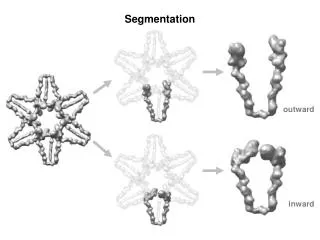
Segmentation outward inward
Three Conformations pre-power stroke state in smooth muscle (PDB - 1BR1) pre-power stroke state in scallop muscle (PDB - 1QVI) Myosin-V with ADP (PDB -1W7I) F-actin
CC= 0.64 CC= 0.58 CC= 0.53 CC= 0.41 CC= 0.42 CC= 0.42 Docking by SITUS (colores) outward inward ADP myosin-V Scallop-like pre-power stroke smooth muscle-like pre-power stroke
CC= 0.64 CC= 0.58 CC= 0.53 CC= 0.41 CC= 0.42 CC= 0.42 Docking by SITUS (colores) outward inward ADP myosin-V Scallop-like pre-power stroke smooth muscle-like pre-power stroke
Docking by SITUS (myoV-ADP) outward CC= 0.64 ADP myosin-V
Normal Mode Flexible Fitting (myoV-ADP) outward CC= 0.82 ADP myosin-V
Docking by SITUS (scallop-like) outward CC= 0.58 Scallop-like pre-power stroke
Normal Mode Flexible Fitting (scallop-like) outward CC= 0.77 Scallop-like pre-power stroke
Fitted “scallop-like” on F-actin pre-power stroke statein smooth muscle myosin (PDB - 1BR1) pre-power stroke statein scallop myosin (PDB -1QVI) Myosin-V with ADP (PDB -1W7I) F-actin F-actin