Download
1 / 204
2.07k likes | 2.21k Vues
Cardiac Anatomy & Physiology. The Heart. Hollow, four chambered, muscular organ. The heart is found in the mediastinum between the right and left lungs. The four chambers are subdivided 2 atria (right & left) 2 ventricles (right & left). Atria.

E N D
The Heart • Hollow, four chambered, muscular organ. • The heart is found in the mediastinum between the right and left lungs. • The four chambers are subdivided • 2 atria (right & left) • 2 ventricles (right & left)
Atria • Each atrium has thin-walls and is separated by the interatrial septum. • The atria act as collecting or holding chambers.
Ventricles • Each ventricle has thick muscular walls and is separated by the interventricular septum. • The ventricles act as pumps. • The right ventricle pumps the unoxygenated blood from your organs and tissues to the lungs. • The left ventricle pumps the oxygenated blood from your heart to your organs and tissues.
Circulation • The vasculature of your lungs is called pulmonary circulation. • The vasculature that supplies the heart with oxygen and nutrients is called coronary circulation. • The vasculature of all of your organs and tissues (everything besides your lungs and heart) is called systemic circulation.
Circulation • The right ventricle is responsible for pulmonary circulation. • The left ventricle is responsible for systemic & coronary circulation.
Valves of the Heart • You have four valves that separate the four chambers of the heart. • Atrioventricular Valves (tricuspid and bicuspid) • Semilunar Valves (pulmonic and aortic valves)
The Valves of the Heart • Atrioventricular Valves (tricuspid and bicuspid) • Semilunar Valves (pulmonic and aortic valves)
Valves • The first valve is between the right atrium & right ventricle. • This valve is called the tricuspid valve. • The valve is called tricuspid because the valve has three flaps. • The flaps are held in place by tendinous cords called chordae tendinae. • The chordae tendinae are secured to the walls of the ventricle by the papillary muscles. • When the ventricles contract the tricuspid valve closes. • When the ventricles relax the tricuspid valve opens.
Valves • The second valve is between the right ventricle and the pulmonary trunk. • This valve is called the pulmonary semilunar. • The valve is called semilunar because of it’s new moon shape. • When the ventricles contract the pulmonary semilunar valve opens. • This allows the blood from the right side of the heart to be pumped to the lungs. • When the ventricles relax the pulmonary semilunar valve closes.
Valves • The third valve is between the left atrium and the left ventricle. • This valve is called the mitral or bicuspid valve. • The valve is called bicuspid because the valve has two flaps. • The two flaps connect to the left ventricle by the same principle as the tricuspid valve. • When the ventricles contract the bicuspid valve closes. • When the ventricles relax the bicuspid valve opens
Valves • The forth valve is between the left ventricle and the aortic trunk. • This valve is called the aortic semilunar. • The valve is called semilunar because of it’s new moon shape. • When the ventricles contract the aortic semilunar valve opens. • This allows the blood from the left side of the heart to be pumped to the body. • When the ventricles relax the aortic semilunar valve closes.
Valves • Valves are suppose to be one-way however they can malfunction. • Valve regurgitation = weak & leaky valve • Valve stenosis = constriction or narrowing of passageway
Valves • Why do valves leak? • Rheumatic fever • Aging • Congenital heart defects
Layers of the Heart • The heart is enclosed in a double walled sac called the pericardium. • It consist of 2 layers • The outer layer is called the fibrous pericardium. • The inner layer is called the serous pericardium. • The serous pericardium consist of 2 layers • Parietal layer • Visceral layer; also called epicardium.
Layers of the Heart • What is Pericarditis? • It is inflammation of the double walled sac called the pericardium. • What causes pericarditis? • Trauma • Infection • Tumors • What are the symptoms? • Chest pain • Audible friction rub
Layers of the Heart • If worsening pericarditis or pericardial effusion can result in cardiac tamponade. • Cardiac tamponade = intrapericardial pressures increase to the point that it impairs the filling of the heart • Cardiac Tamponade is life threatening and is sometimes treated with pericardiocentesis.
Layers of the heart • The wall of the heart is made up of three layers. • Epicardium • Corresponds to the visceral pericardium. • Functions as an outer protective layer. • Serous membrane that consists of connective tissue covered by epithelium. • Includes blood capillaries, lymph capillaries, and nerve fibers.
Layers of the heart • The wall of the heart is made up of three layers. • Myocardium • Relatively thick. • Consists largely of cardiac muscle tissue responsible for forcing blood out of the heart chambers. • Muscle fibers are arranged in planes, separated by connective tissues that are richly supplied with blood capillaries, and nerve fibers.
Layers of the heart • The wall of the heart is made up of three layers. • Endocardium • Consists of epithelial and connective tissue that contains many elastic and collagenous fibers. • Connective tissue also contains blood vessels and some specialized cardiacmuscle fibers called Purkinje fibers. • Lines all of the heart chambers and covers heart valves. • Is continuous with the inner lining of blood vessels--endothelium.
Wall of the Heart • What is endocarditis? • It is an infection and inflammation of the heart's inner lining (endocardium). It is most common in people with damaged, diseased, or artificial heart valves. • What causes it? • It is caused by bacteria that enter the bloodstream and settle on the heart valves. • What are the symptoms? • Chills & Fever • Fatigue • Weight loss • Painful joints • Persistent cough and SOB • How is it treated? • IV Antibiotics
Blood supply to the Heart • The two main arteries that feed the heart: • Left coronary artery • Circumflex branch • Anterior interventricular branch • Right coronary artery • Marginal branch • Posterior interventricular branch
Blood supply to the Heart • The main veins that drain “used” blood from the heart: • Great cardiac veins • drains the anterior side of the heart • Middle cardiac vein • Drains the posterior side of the heart • The great cardiac and middle veins merge together into a cavity called the coronary sinus. • The thebesian vein then carries the “used” blood into the left and right atria.
Disorders • Atherosclerosis = hardening of the arteries which promotes clots and/or occlusions. • Thrombosis = a clot /coagulation of blood • Embolism = thrombosis that has traveled from location it was formed. • Myocardial Ischemia = decreased oxygen availability to the heart because of decreased blood flow or decreased oxygen in blood. • Myocardial Infarction = tissue death due to a loss of blood & glucose to the heart muscle.
Disorders • Congestive Heart Failure (CHF) = condition where the left side of the heart is damaged. • Cor Pulmonale = condition where the right side of the heart has decreased function. • Angina Pectoris = a severe pain or pressure in the chest caused by inadequate blood flow and oxygen content to the heart muscle.
Treatment for Disorders • Coronary Angioplasty = treats blockages of vasculature with a catheter or balloon. • Coronary Artery Bypass Graft (CABG) = artery graft from the leg or arm is inserted into coronary vasculature to bypass blocked arteries.
Blood flow through the Heart • Inferior Vena Cava/ Superior Vena Cava • Right Atrium • Tricuspid Valve • Right Ventricle • Pulmonary Semilunar Valve • Pulmonary artery trunk • Pulmonary artery • Left/Right pulmonary artery • Lungs
Blood flow through the Heart • Left/Right pulmonary vein • Left Atrium • Bicuspid/Mitral Valve • Left Ventricle • Aortic Semilunar Valve • Aortic artery trunk • Ascending Aorta • Brachiocephalic artery • Left common carotid artery • Left Subclavian artery • Descending Aorta • xxxxxxxxxxxxxxxxxxxxxxxxxxxxxxxxxxxxxxxxxxxxxxxxxx
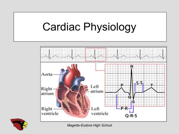
Cardiac Cycle • Cardiac cycle is the term referring to all or any of the events related to the flow of blood that occur from the beginning of one heartbeat to the beginning of the next
The frequency of the cardiac cycle is the heart rate Every single 'beat' of the heart involves three major stages: atrial systole ventricular systole complete cardiac diastole The term diastole is synonymous with relaxation of a muscle. It is the period of time when the heart relaxes after contraction in preparation for refilling with circulating blood.
The term systole is synonymous with contraction (movement or stretching) of a muscle. Think squeeze The term diastole is synonymous with relaxation of a muscle. Think dilate. It is the period of time when the heart relaxes after contraction in preparation for refilling with circulating blood.
Heart Rate • Heart rate is a term used to describe the frequency of the cardiac cycle. • It is considered one of the four vital signs • Usually it is calculated as the number of contractions (heart beats) of the heart in one minute and expressed as "beats per minute" (bpm). • Normal Heart rate in adults 60-100 bpm
Stroke Volume • Stroke volume is the amount of blood pumped by the left ventricle of the heart in one contraction • The heart does not pump all the blood out of the ventricle. Normally, only about two-thirds of the blood in the ventricle is put out with each beat • Normal range • 60 -120mL
Cardiac Output (Qt) • Cardiac output is the volume of blood being pumped by the heart, in particular a ventricle in a minute. • Cardiac Output (CO) = SV × HR • Normal range is 4-6 lpm
Electrophysiology of the Heart • Contraction of the heart is initiated by an electrical stimulus • These contractions are a function of action potentials (electrical currents) • Action potentials consist of 5 phases • 0 = depolarization • 1-4 represent polarization