Download
1 / 12
130 likes | 319 Vues
Layers of the Retina. Horacio M. Tous, M.D Department of Ophthalmology University of Puerto Rico. Layers of the Retina. The Internal Limiting Membrane (ILM).

E N D
Layers of the Retina Horacio M. Tous, M.D Department of Ophthalmology University of Puerto Rico
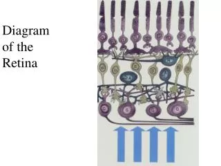
The Internal Limiting Membrane (ILM) The innermost layer is the place of fused Muller cells foot processes. This fusion completes the function of sealing off the retinal components from the potentially harmful materials present in the vitreous chamber. It is a region of occluding junctions and a "tight" barrier.
The Nerve Fiber Layer (NFL) The unmyelinated ganglion cell axons bundled together, run radially around the inner surface of the retina, converging at the site of origin of the optic nerve. Myelination by oligodendrites occurs at the lamina cribosa.
The Ganglion Cell Layer (GCL) It’s where the ganglion cell bodies are. The axons from these cells are bundled into tracts that run radially, forming the next layer of the retina.
The Inner Plexiform Layer (IPL) The inner plexiform layer is a region of synapses. Here the bipolar cell processes synapse with the dendritic processes of ganglion cells, as well as amacrine cells.
The Inner Nuclear Layer (INL) It’s where the cell bodies and nuclei of the bipolar, amacrine, muller and horizontal cells are located.
The Outer Plexiform Layer (OPL) This is the site of numerous synapses between the rod and cone cells and the dendritic, processes of horizontal, amacrine and bipolar cells. Middle limiting membrane forms approximate border of vascular inner portion and avascular outer portion of the retina. Central retinal artery supplies retina internally(MLM toILM) Choriocapillaris supplies retina externally (MLM to RPE)
The Outer Nuclear Layer (ONL) This layer is the location for the nuclei and cell bodies of the rod and cone cells.
The Outer Limiting Membrane (OLM) It’s the site of numerous occluding tight junctions. The junctions are between the plasma membranes of the rod segments and the Muller cells. They serve to isolate the inner layers of the retina from potentially harmful material in the blood circulation, forming a blood-retina barrier.
Layer Rods and Cones (R&CL) The outermost layer of the retina. These are the actual light-sensitive elements.
Pigment Epithelium Layer (PE) It is a single layer of cuboidal cells. These cells are impregnated with melanin, but they also contain lipofuscin as a product of their metabolic activity. The pigment layer cells phagocytose the ends of the rods and cones as they are renewed: hence the accumulation of lipofuscin. Another and even more important function of the pigment epithelium is the storage and synthesis of trans-retinal, or vitamin A.