Different Haemostatic Techniques in Laparoscopic surgery
610 likes | 1.4k Vues
Different Haemostatic Techniques in Laparoscopic surgery. by: Dr/ Adel Fathi lecturer of surgical oncology – OCMU - Egypt (Oncology Center Mansoura University). As in open surgery, uncontrolled bleeding during laparoscopy is one of the major surgical pitfalls.

Different Haemostatic Techniques in Laparoscopic surgery
E N D
Presentation Transcript
Different Haemostatic Techniques in Laparoscopic surgery by: Dr/ Adel Fathilecturer of surgical oncology – OCMU - Egypt(Oncology Center Mansoura University)
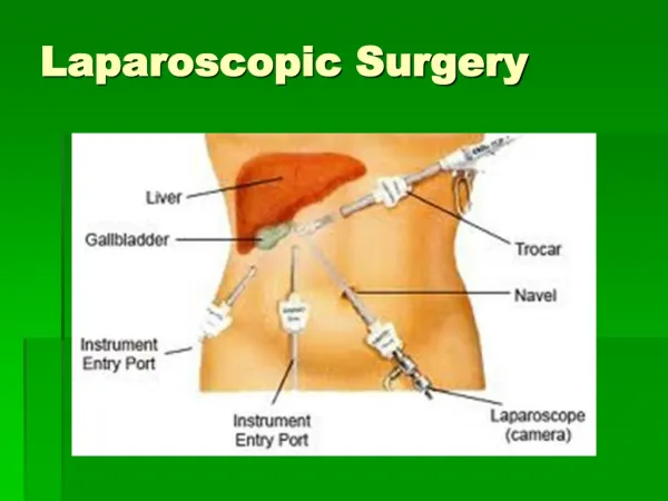
As in open surgery, uncontrolled bleeding during laparoscopy is one of the major surgical pitfalls. Haemorrhage may occur by gaining laparoscopic access, during surgical preparation or during ablative and reconstructive surgery. In addition, bleeding may interfere with vision during laparoscopy owing to significant light absorption by dark blood staining of the adjacent tissue within the magnified optical field during laparoscopy. Introduction
Dissection with a sponge or haemostatic stick helps to dissect tissue and control blood vessels locally. Local compression with a sponge is similar to digital compression in open surgery, especially in venous bleeding, and gives the surgeon time for further strategies for final haemostasis, or it may be sufficient alone.
Intracorporial suturing is preferable than external knotting as it avoids excessive traction during suturing
Disadvantages of suture technique: 1- Suturing requires advanced laparoscopic skills. 2- A significant amount of healthy tissue needs to be sacrificed. 3- Loops may slip off owing to tissue ischemia with secondary loosening of the fixed loops.
Clip system is the preferable method of sealing blood vessels as suturing requires advanced skills. • Small ……. 6mm • Medium/large…… 9mm • Large ……… 10-11mm
Disadvantages of clip technique: 1.The reloading time of single fire instruments is an important limitation in emergency situations. 2.Titanium clips tend to slip off during further dissection. 3.At least 2 to 5 clips seem mandatory for safe control of vessels sized ≥ 3mm.
1- The force required to dislodge the absorbable clip was significantly greater than for metallic clips 2- The adhesion scores tended to be lower with the absorbable clips 3- Titanium clips produce rarefaction in C.T. but it is used for radiographic markings ## Absorbable polymeric clips were hemostatically effective in this laparoscopic model and may offer advantages over metallic clips (Klein et al., 1994) Comparison of titanium and absorbable polymeric surgical clips:
Vascular Endo-stapler (vascular Endo-GIA) with 2.0 to 2.5 mm jaw width and various lengths have been used to achieve safe occlusion of major vessels or vascular pedicles. The major disadvantage of endo-staplers may occur when major vessels are sealed insufficiently, resulting in life-threatening bleeding.
As in open surgery, a monopolar current is used widely for haemostasis and was the first tool adapted for laparoscopic needs. Predominant instruments used for monopolar dissection or cutting are forceps, scissors or the J-hookelectrocautery, attached to a monopolar generator.
1.Large amount of smoke and steam 2.They predispose surgical wounds to postoperative infection. 3.Electrosurgery devitalizes tissues at the wound edges. 4.The use of the electrosurgical tools causes worse cosmesis at closure. 5. Incidence of burn due to insulation failure or direct coupling or capacitive coupling injuries Disadvantages of monopolar
The appearance of burns are common following Monopolar in laparoscopy; particularly at the port site of the active electrode. Willson et al (1997) found that 9 out of 19 skin biopsies from the skin adjacent to the port site of the monopolar instrument's active electrode were found to have thermal injury by histology. Thermal injuries
A- insulation failure Defects in the insulation, too small to be visually perceptible, may deliver current outside the field of view
B- Direct coupling Direct coupling can transfer 100% of the current to bowel outside the field of view when a plastic cannula is used around the laparoscope
Anytime two conductors are separated by an insulator a capacitor is created. For example, a capacitor is created by inserting an active electrode, surrounded by its insulation, down a metal cannula C- capacitive coupling
Capacitively coupled electrical current can be transferred from the active electrode, through intact insulation and into the conductive metal cannula. Then the cannula come in contact with body structures, that energy can be discharged into these structures and damage them. With an all-metal cannula, electrical energy stored in the cannula will tend to disperse into the patient through the relatively large contact area between the cannula and the body wall. Because the contact area is relatively large, the electrical energy is less concentrated and less dangerous. For this reason it is unwise to use plastic anchors to secure the cannula because it isolates the current from the body wall.
Some institutions use plastic trocarcannula systems because they wrongly believe them to be safer. The plastic systems can also be dangerous because the patient’s own conductive tissue within the body can form the second conductor, creating a capacitor. The patient’s omentum or bowel draped over the plastic cannula could discharge stored energy to adjacent body structures.
A "capacitance injury" may occur as a result of induced currents through intact insulation when a hybrid cannula (metal cannula with plastic gripper) is used
• Inspect insulation carefully • Use lowest possible power setting • Use low voltage (cut) waveform • Use brief intermittent activation vs. prolonged activation • Do not activate in open circuit • Do not activate electrode in close proximity or direct contact with metal/conductive object • Use bipolar electrosurgery when appropriate • In the operative channel for activated electrodes - Select an all metal cannula system as the safest choice - Do not use a hybrid system (metal and plastic components) • Utilize available technology: - Tissue response generator to reduce capacitive coupling in the low voltage waveform - Active electrode monitoring system to eliminate concerns with insulation failure and capacitive coupling Recommendations to avoid electrosugical thermal injury during minimally invasive surgery are:
Bipolar instruments resemble surgical forceps, with both the active electrode and the return electrode functions being performed at the surgical site. The electrosurgical energy does not travel through the patient but is confined to the tissue between the forceps. Introduction of bipolar instruments in laparoscopy improved safe dissection and haemostasis simultaneously, thus allowing early vascular and bleeding control while avoiding complications.
LigaSureTM is the most advanced system which was released in Japan in 2000. • It has been used widely in open and laparoscopic surgery and consists of a bipolar radio frequency generator and a laparoscopic Maryland style forceps.
It is a new hemostatic system based on pressure and bipolar electrical energy, which seals vessels as large as 7 mm in diameter. • The device delivers a controlled high-power current at a low voltage to melt collagen and elastin, permanently fusing the vascular layers and obliterating the vessel lumen.
The harmonic Scalpel is a cutting instrument used during surgical procedure to simultaneously cuts and coagulates tissue. Harmonic Scalpel The device itself is composed of a generator, hand piece and a blade.
Electrical energy is transferred from a microprocessor controlled generator to a transducer in the hand piece. The generator pulses the transducer with Alternating Current (AC) at its natural frequency of 55,500 Hz. Mechanism of action
1. less smoke production and less thermal energy spread. 2. It has low temperature vaporization of tissue 3. Harmonic Scalpel simultaneously cuts or dissects tissue. 4. Since no electrical current is passed into or through the patient, there is no risk of direct coupling or capacitive coupling injuries. 5. There is also no risk of neuromuscular stimulation. This is especially important in patients with pacemakers or implanted defibrillators. Advantages of the Harmonic Scalpel
1. The functioning blade is hot and will remain so seconds after an application. Contact with bowel or other surrounding vital structures with the functioning blade should be avoided at all times. 2. There are also occasional reported events of the distal tip of the instrument falling off during surgery. 3. Failure of the device to test properly before activation at the beginning of the case. Disadvantages of Harmonic Scalpel
4. There are a few reported instances of the blades becoming overheated and melting trocars during certain laparoscopic procedures. 5. Some studies showed how many viable cancer and blood cells are present in the aerosols produced by the Harmonic Scalpel. 6. Proper training and experience with the device is necessary to prevent inadvertent damage to surrounding structures.
Lasers were introduced in to the medical field during the 1960s, and currently a number of surgical lasers are available with more superseded by more practical technologies. CO2-, Argon, and Nd:YAG lasers are being used. The effect of laser is primarily thermal.
The first haemostatic device used for capillary bleeding control. This instrument uses an argon jet to blow off the surgical field and is sufficient for minor capillary bleeding after dissection. Argon beam coagulator
1. Can not be used for tissue dissection and is unsuitable for control of significant bleeding or larger vessels. 2. Argon gas embolism or pneumothorax have occurred even when used correctly. Disadvantages of Argon beam
These devices are primarily used to dissect parenchymatous organs or to perform the total mesorectal excision. They help to identify vessels and bile ducts that can be closed by clips or ligature.
CUSA (Cavitron Ultrasonic Surgical Aspirator) is an ultrasonic-dissection system that works with mechanical energy. A continuous suction system is integrated, which keeps the tissue close to the oscillating tip of the instrument to increase the efficiency and to clean the operating area.
HYDRO-JET® uses a fine laminar jet of water on its surface drilled that leads to a highly differentiated mode of application and tissue selection.