Pediatric endodontic materials
Department of Pediatric & Preventive Dentistry. Pediatric endodontic materials. Presented By: Nilesh Deshpande , Junior Resident-II. Contents. Introduction Definition Different Obturating materials Recent advances Conclusion Bibliography. Introduction.

Pediatric endodontic materials
E N D
Presentation Transcript
Department of Pediatric & Preventive Dentistry Pediatric endodontic materials Presented By: NileshDeshpande, Junior Resident-II.
Contents • Introduction • Definition • Different Obturating materials • Recent advances • Conclusion • Bibliography
Introduction • The final of endodontic treatment is to fill the entire root canal system and all its complex anatomic pathways completely and closely with non irritating hermatic sealing agents. • Obturation of a root canal should result in a complete seal from the coronal aspect to the apex preventing the entry of microorganisms and fluid
Pulpectomy • Mathewson in 1995 defined it as the complete removal of necrotic pulp from the root canals of primary teeth and filling them with an inert restorable material so as to maintain the tooth in the dental arch. • Finn defines it as removal of all pulpal tissues from the coronal and radicular portions of the tooth.
Ideal Requisites (by Castagnola et al): • The material should resorb as the roots of primary teeth resorbs. • Should not irritate periapical tissues. • Should not coagulate any organic remnants in the canal. • Should be able to adequately disinfect and seal the canals. • Should be non toxic. • Should not get dissolve in oral fluids. • If surplus material has been filled in periapical area then it should be easily resorbed.
Should have proper consistency on mixing so that it can be adequately pushed into the canal. • It should not discolour the tooth. • Should be radiopaque. • Should be retrievable if required. • Should be harmless to the adjacent tooth bud.
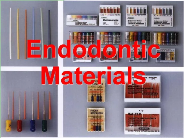
Materials used for Obturation: Primary Teeth Permanent Teeth - Zinc oxide eugenol - Guttapercha - Ca(OH2) - Silver Cones - Iodoformpaste - Stainless Steel File method
Commercial Products Available... B. KRI paste: • Iodoform 80% • Camphor 4.86% • Parachlorophenol 2.025% • Menthol 1.215% A. Walkhoff Paste: • Parachlorophenol • Camphor • Phenol C. Vitapex / Metapex: • Calcium Hydroxide • Iodoform • Oily Additives E. Maisto Paste: • ZnO • Iodoform • Thymol • Chlorophenol • Lenolin D. Endoflas: • Barium Sulphate • Calcium Hydroxide • Iodoform • ZnOE
ZINC OXIDE EUGENOL PASTE • It is the most frequently used oburant. • The filling material of choice is ZnOE without a catalyst. • Lack of catalyst gives adequate working time for filling the canals.
NOTE: The most popular of all root canal filling material for primary teeth are ZnO and eugenol (ZnOE), calcium hydroxide [Ca(OH)2] and iodoform pastes. • Both ZnOE & Metapex/ Vitapex have shown encouraging results.
Review of Literature • Few studies have reported that ZnOE sets into a harder cement that resists resorption when extruded beyond the apices. • Investigation by Mani et al reported 67% of all overfilled canals showed over-retained ZnOE at 6 months follow-up. • Flaitz et al1964reported that 20% of the permanent teeth showed deflection in case of overfilling of canals with ZnOE. • Eransqun & Munuzabul1972 reported that ZnOE irritates the periapical tissue of rats and may produce necrosis of bone and cementum.
Months/years to resorb Grossman, 1974 woods, 1984 • Jevell& Ronk1982 reported that premolar eruption was arrested due to toxic effects of ZnOE. • According to Reddy et al1985lack of ZnOE’s antibacterial properties may aggravate residual infection of root canal instead of promoting healing of infected tissue. • Coll et al1992 reported it has no significant effect on exfoliation of primary teeth in any of the case.
Goeriget al in their study reported that ZnOE when mixed with other root canal filling materials like calcium hydroxide & iodoform has good antimicrobial activity and resorption capabilities as when used alone. • In 1985 Collreported that ZnOE could alter the path of eruption of succedneous teeth. • Wright KJ 1994 did a study on comparison and antimicrobial effects of ZnOE & KRI paste (ZnOE+Iodoform). Results suggested that ZnOE has better antimicrobial activity then KRI paste. It had lower cytotoxicity.
Coll et al1998in their study reported that gross own filling was related to failure of pulpectomy with ZnOE. • According to Sadiuan et al2000 tooth overfilled with ZnOE and those filled up to the apex did not show resorption as compared to those which were filled 1 mm or short of the apex. • Holan et al2001 reported that 100% of the tooth filled to the apex with KRI paste and 85% of those filled with ZnOE were successful and not statistically different.
No difference was observed when the teeth were under filled with ZnOE or KRI paste. • Overfilling of canals, however, resulted in a much higher success rate of KRI (75%) then ZnOE (41%), which was statistically significant difference.
Advantages: • Easily available. • Radiopaque material. • Cheaper/ cost effective. • Effective antimicrobial agent. • Also less cytotoxic to cells in direct or indirect contact. • Good plasticity • Insoluble in tissue fluids
Disadvantages: • Excessive filling - it leads to mild foreign body reaction. • Muruzabul found that ZnOEcement was “highly irritating” to the periradiculartissues and caused necrosis of bone and cementum. • Rate of resorption does not coincide with rate of resorption of root ( a little slow).
Coll and Sadrianreported that ZnOE retained material alter the path of eruption of succedeneous teeth in 20% of cases. (However it has been shown that optimally filled and overfilled canals showed a statistically higher success rate compared to underfilled root canals). • It has been found that Eugenol is not only cytotoxic but is neurotoxic also.
NOTE: • Canals are said to be underfilled when the material is filled 2 mm short of radiographic apex. • Lentulo-spiral if chosen for obturation then it should be smaller by 2 size from the last H/K file used and 1mm short of working length.
Composition of ZnOE Cement Consists of: • Powder: ZnO+ Staybelite resin+ Bismuth subcarbonate + Sodium borate + BaSO4 • Liquid : Eugenol Note: At the time of placement pH of cement is 7 which potentially makes them as the least irritating of all dental materials.
Manipulation • It consists of powder containing ZnO and liquid containing Eugenol. • Powder contains finely ground ZnO which enhances flow of cement. • It has been shown that 1 mm of ZnO Eugenol cement has a radiopacity corresponding to 4.5 mm of Aluminum sheet, which is slightly lower than G.P.
Resin acids (Monobasic carboxylic acids) when mixed with ZnOE it renders it less soluble than regular ZnOE cement. • Consistency of paste when used for filling should be 1 scoop powder : 1 drop liquid. • Consistency for temporary filling ZnOE cement should be 2:1.
CALCIUM HYDROXIDE • Ca(OH)2 a colourless crystal or white powder. • Prepared by reacting Ca oxide (lime) with water, a process called slaking and is also known as hydrated lime or slaked lime. • When heated above 5800C it dehydrates forming the oxide. • Hermann introduced Ca(OH)2 in endodontics in 1930.
PROPERTIES OF Ca(OH)2 1)Antibacterial action 2)Heals peri apical lesions 3)Resorbs easily 4)Does not set intohard mass 5)No discoloration of teeth
Review of Literature • In 1938, Teuscher and Zander introduced the rationale that Ca(OH)2 has ability to form reparative dentin. • Seltzer and Benden identified the osteogenic potential of Ca(OH)2. • Tanburic et al summarized the mineralizing effects of Ca(OH)2. • Estrela et al summarized the antibacterial properties of Ca(OH)2
There was some controversy regarding the source of Calcium ions in the dentin bridge repair at exposure site: • Sciaky and Pisanti, Attalla and Norjain demonstrated that Ca ions from capping materials were not involved in bridge formation. • Stark and his colleagues, however believed that Ca ions come from the blood circulation which they have shown by presence of radiolabelled Ca ions.
Tuan T.J. in 1957, reported that Ca(OH)2 could inhibit macrophage function and reduces inflammatory reaction in periapical tissue or in pulp when it is used in pulpectomy or in DPC.
Composition of Ca(OH)2 Sealer Base PasteCatalyst Paste • Glycol salicylate (40%) - Ca(OH)2 (50% ) • Ca(SO4)2 - ZnO (10%) • Titanium dioxide (Inert filler) - Zn stearate (0.55%); accelerator • Calcium tungstate or Basulphate - Ethyl toluene (Provides radiopacity) - Sulphonamide (39.5%); oily compound, acts as a carrier
Advantages • Has got an antibacterial action (Initially bactericidal than bacteriostatic) : • Hydrolyses bacterial cell wall lipopolysaccharides thus making them incapable of producing biologic effects such as toxicity, pyrogenecity and complement activation. • Neutralizes bacterial endotoxins. • Reduces anaerobic organisms through CO2 absorption.
Protection of pulp (If used in pulpotomy procedure) • Is an ideal pulp protection agent. • If used beneath acid containing bases/cements, it neutralizes the acid due to its high alkalinity. • Should be used in a very thin layer over or near pulp exposures. • Obliterating the canal space with Ca(OH)2 during treatment may minimize the ingress of tissue fluid used as a nutrient by microorganisms.
Blocks patent dentinal tubules, neutralizing attack of inorganic acids and leached products from certain cements and filling materials. • Promotes healing and repair.
Disadvantages • Pulp Obliteration: • Due to osteogenic potential, it is capable of inducing calcific metamorphosis, resulting in obliteration of pulp chamber and root canals.
Internal resorption: • Induces internal resorption in primary teeth. • However investigations have reported that mixture of Ca(OH)2 and iodoform (Metapex/vitapex) is easy to apply, resorbs at a slightly faster rate than roots and has no toxic effects on the permanent successor and is radiopaque also.
Internal resorption results due to overstimulation of Undifferentiated mesenchymal cells leading to formation of odontoclasts. These odontoclasts then resorb the dentin. • Although it has been documented that internal resorption occurs due to Ca(OH)2 in primary teeth, it does not appear to be a problem in permanent teeth.
Lack of adhesion to hard tissue: • It is a major shortcoming of Ca(OH)2. • This leads to inadequate seal against microleakage. • Furthermore, Ca(OH)2 materials have been found to dissolve under restorations where microleakagehas occurred, resulting in bacterial access to the pulp. • Does not adhere to dentin or resin restoration.
IODOFORM PASTE Review of Literature: • Castagnola and Urley in 1952 demonstrated that KRI paste got resorbed with root at the same rate which was seen as a success. • It is bactericidal to microorganism in root canals and loses only 20% of potency over a 10 year period. • Maistoin 1967 introduced a mixture of ZnOE and Iodoform (Maisto paste) as a RC filling material in permanent teeth, but was used by Tagger and Sarnet in 1984 in primary teeth.
Garcia Godoy in 1987 found that KRI paste is bactericidal in root canals, resorbs from the apical tissues in one or two weeks, is apparently harmless to tooth germs, is radiopaque, does not get to a hard mass and is easily inserted and removed. • Mass in 1989 found Maisto’s paste to be successful in treating an infected primary posterior teeth. He reported that iodoform containing pastes are easily resorbed from the periradicular region and causes no foreign body reaction like ZnOE.
Doninguezin 1989 reported that when iodoform and Ca(OH)2 are combined excellent clinical, radiographic and histological results were obtained. • Matsuzuki in 1996 reported that iodoform improved antiseptic and radiographic effects. • Reddy and Fernandes in 1996 found Maisto paste to be 100% successful. 93% should bone regeneration with complete healing of interradicular pathology and complete resorption of excess material.
Nucko and Godoy in 1999 evaluated effectiveness of vitapex Ca(OH)2 +iodoform paste and found that it is radiopaque, does not set to a hard mass, resorbs from the apical tissues in 1 week to 2 mos., apparently harmless to permanent tooth germ and can be easily inserted and removed. • Chawla HS in 2001 evaluated the effect of mixture of ZnO powder, Ca(OH)2 paste and distilled water as a root canal filling material. They found that material resorbed at the same rate as the root.
Fuks et al 2002 conducted a study using endoflas as filling material. After 52 months, overfilling led to a success rate of 58%, underfilling showed a success rate of 83%. The paste also got resorbed extra radicularly. • In a study conducted on 96 primary molars for clinical and radiographic evaluation of pupectomies using ZnOE and iodoform (RC fill), Ca(OH)2 and iodoform (vitapex), ZnOE+ Ca(OH)2 +Iodoform (endoflas) in 72 children of age group 4-7 years, it was reported that success rate was: 90.6% with Metapex 84.7% with RCfill 95.1% with Endoflas
IODOFORM • It is preparation of iodine. • Obtained by the action of chlorinated lime upon an alcoholic solution of iodide of potassium heated at 1040 deg F. • The product being iodoform and iodate of lime. • Iodoform has no irritant action. • In small doses it relives pain, disinfectant having great influence on the nervous system.
KRI paste • Highly resorbable, bacteriocidal. • Iodoform 80% • Parachlorophenol 2%, • Camphor 5%, • Menthol 1%. • Fuks et al 2000 found that • Success rates of 84% with the Kri paste group versus 65% with the ZOE group. • Overfills more successful (Kri paste 79% vs. ZOE 41%). The excess paste will resorb without causing any adverse side effects.
WALKHOFF PASTE Composition • Camphorated Parachlorophenol : 4-8% • Eugenol: 24 to 22% • Zinc oxide: 48 to 58% • Di-iodothymol: 12 to 18% • Menthol crystals: 1.40 to 2.90% • Silver powder: 0.70 to 1.45% approx.
Parachlorophenol • Antiseptic agent • Dissolve albumins and which can therefore progressively penetrate into the canaliculi of the tooth. • Major disadvantage is its total resorption, which occurs both in the periapical area and in the canal area of the tooth. • Even in the most favourable cases, there is no longer any trace of paste in the previously filled canals, one year after filling of the canals has taken place.
Camphor • To allay the pain arising from the near exposure of the pulps of teeth • Also the pain of sensitive dentine • Also to arrest the hemorrhage and allay the pain of wounded pulps of teeth.
Menthol • Anodyne • Antispasmodic • Antiseptic • Menthol has given satisfaction as an external remedy in facial neuralgia, odontalgia, as an obtunder of sensitive dentine, and as a local anesthetic
ENDOFLAS • Endoflas is root canal sealer material • Composed of zinc oxide,bariumsulfate,iodoform,calciumhydroxide,eugenol and pentchlorophenol • One condition for success of endoflas is prevention of microleakage. • A permanent restoration should be placed as soon as possible after clinical signs and symptoms of inflammation are eliminated.
Chawla H.S et al in 2008 performed endodontic treatment on 25 pulpally involved mandibular primary molars in 4-9 yrs old children, the root canals were obturated with new root canal filling material consisting of mixture of calcium hydroxide ,zinc oxide and 10% sodium fluoride solution. • All cases were clinically and radio graphically evaluated after 3 months and 6 months. • It was observed that the rate of resorption of this new root canal obturating mixture was quite similar to rate of physiologic root resorption in primary teeth.