Download
1 / 1
70 likes | 686 Vues
University “ Goce Delcev ” Shtip , Faculty of Medical Sciences Department of Stomatology*. University “St. Kiril and Metodij ” Skopje*** Faculty of Dentistry l Private dental clinic “ Denkovski ”: ** R . Macedonia. OUR EXPERIENCE WITH THE Nd : Yag LASER IN THE THERAPY

E N D
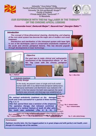
University “ GoceDelcev” Shtip, Faculty of Medical Sciences Department of Stomatology* University “St. Kiril and Metodij” Skopje*** Faculty of Dentistry l Private dental clinic “ Denkovski”: ** R. Macedonia OUR EXPERIENCE WITH THE Nd: Yag LASER IN THE THERAPY OF THE CHRONIC APICAL LESIONS Kovacevska Ivona*, DenkovskiMarjan**, Nacevski Ivan**,Georgiev Zlatko***. Introduction The concept of three-dimensional cleaning, disinfecting, and shaping of the root canal system has become the major aim of modern root canal treatment. Disinfection and sterilization of the intracanal system with laser light is one of the techniques used in conservative endodontic treatment of the acute and chronic periapical lesions. This has become popular through developments in laser technology. Objective Our goal was to make clinical and radiographic assessment of the therapeutical effects of the Nd: Yag Laser with the chronic periapical lesions. Fig. 1 – Fidelis lll Fig. 2 – Micro fiber endodontic treatment Study design In the study, we present cases of single and multi-rooted human teeth with X-ray determined periapicalradiolucency. Intracanalsterilization and disinfection was realized in 56 teeth. In the first session the teeth were opened, the canal contents was removed, and drainage was established so that the symptoms are calmed. During the second visit we determined the canal length with a #10K file at 1mm from the apex. The root canals were instrumented with the crown-down technique up to #40K, or #45K file depending on the canal’s magnitude. Two ml 1% sodium hypochlorite was used between each file change. After instrumentation a final irrigation with 10 ml of distilled water was performed. The canals were then dried with paper points. The optic fiber, 300μm wide, from the pulsed Nd YAG laser was set in each canal and laser radiation with circular slow motions from the apex to the crown was applied 3 times in 5 seconds period. We realised endodontic treatment on the damaged tooth and it was succesfull for a period of three months. (Fig. 5, 6, 7) After that period there was a remision of the simptoms. The operative therapy that followed confirmed a presence of a vertical fracture af tooth 24 after which the tooth was extracted. (Fig. 8, 9, 10, 11) Fig. 4 - intraoral status Fig. 3 - intraoral status Fig. 6 - root canal preparation Fig. 5 - extirpation Fig. 9 and 10 extracted tooth 24 Fig. 1 Fig. 8 - operative access Fig. 7 - temporary filling Fig. 11 - after radical therapy Conclusion: Eighteen months later, the four-legged patient is in great shape and with perfect oral health, even though it’s missing one of its canines.