Maximizing Genetic Information
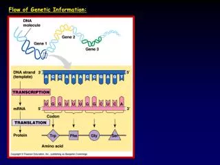
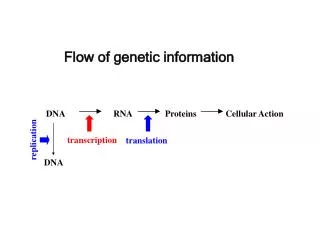
The human genome contains about 20,325 genes - However, these encode about 100,000 mRNAs, which in turn specify more than a million proteins Several events account for the fact that proteins outnumber genes. Maximizing Genetic Information. Maximizing Genetic Information. Figure 11.8.

Maximizing Genetic Information
E N D
Presentation Transcript
The human genome contains about 20,325 genes - However, these encode about 100,000 mRNAs, which in turn specify more than a million proteins Several events account for the fact that proteins outnumber genes Maximizing Genetic Information
Maximizing Genetic Information Figure 11.8 Figure 11.11
The “genes in pieces” pattern of exons and introns and alternate splicing help to greatly expand the gene number Maximizing Genetic Information Figure 11.9
Only 1.5% of human DNA encodes protein Rest of genome includes: - Viral DNA - Noncoding RNAs - Introns - Promoters and other control sequences - Repeated sequences Most of the Human GenomeDoes Not Encode Protein
About 8% of our genome is derived from RNA viruses called retroviruses - This is evidence of past infection - Sequences tend to increase over time Viral DNA Figure 11.11
RNA • sncRNA: stands for small noncoding RNA also called microRNA ( miRNA,miRs).. typically composed of only 22 nucleotides, and acts as a negative regulator of genes ( meaning they turn the genes off) • sncRNA controls 20-30% of human coding DNA genes ( whole mechanism is called RNAi or RNA interference)
Nearly all of the human genome can be transcribed, and much of it is in the form of noncoding RNAs (ncRNAs) This includes rRNAs and tRNAs However, there are hundreds of thousands of other ncRNAs - These are transcribed from pseudogenes - But are not translated into protein Noncoding RNAs
Transposons are the most abundant type of repeat - Sequences that jump about the genome - Alu repeats can copy themselves - Comprise about 2-3% of the genome Rarer classes of repeats include those that comprise telomeres, centromeres, and rRNA gene clusters Repeats
The Nature of Mutations A mutation is change in a DNA sequence that is present in < 1% of a population May occur at the DNA or chromosome level A polymorphism is a genetic change that is present in > 1% of a population The effect of mutations vary “Loss-of-function” mutations – Recessive “Gain-of-function” mutations – Dominant
The Nature of Mutations The term mutant refers to phenotype - Usually connotes an abnormal or unusual, or even uncommon variant that is nevertheless “normal”
The Nature of Mutations Germline mutations - Originate in meiosis - Affect all cells of an individual Somatic mutations - Originate in mitosis - Affect only cells that descend from changed cell
Mutations Alter Proteins Identifying how a mutation causes symptoms has clinical applications Examples of mutations that cause disease: - Beta globin gene - Collagen genes
Sickle Cell Anemia Results from a single DNA base change in the b-globin gene, which replaces glutamic acid (6th position) with valine Phenotype associated with homozygotes Altered surface of hemoglobin allows molecules to link in low oxygen conditions Creates sickle shape of RBC Sickling causes anemia, joint pain, and organ damage when RBC become lodged in small blood vessels
Thalassemia Caused by another beta hemoglobin mutation Too few beta globin chains Excess of alpha globin prevents formation of hemoglobin molecules So RBCs die Liberated iron slowly damages heart, liver, and endocrine glands Thalassemia minor (heterozygous) Thalassemia major (homozygous for mutation and more severe)
Collagen A major component of connective tissue - Bone, cartilage, skin, ligament, tendon, and tooth dentin More than 35 collagen genes encode more than 20 types of collagen molecules Mutations in these genes lead to a variety of medical problems
Collagen has a precise structure - Triple helix of two a1 and one a2 polypeptides - Longer precursor, procollagen is trimmed to form collagen Figure 12.3
Figure 12.4 Ehler-Danos Syndrome A mutation prevents procollagen chains from being cut Collagen molecules cannot assemble, and so skin becomes stretchy Figure 12.4
How Mutations Cause Disease Mutations in a gene may cause either different versions of the same disease or distinct illnesses Table 12.2 lists several examples of mutations and the diseases they produce Figure 12.4
Causes of Mutations Mutations may occur spontaneously or by exposure to a chemical or radiation An agent that causes a mutation is called a mutagen Figure 12.4
Spontaneous Mutation De novo or new mutations Not caused by exposure to known mutagen Result from errors in DNA replication DNA bases have slight chemical instability Exist in alternating forms called tautomers As replication fork encounters unstable tautomers, mispairing can occur Figure 12.4
Spontaneous Mutation Rate Rate differs between genes - Larger genes usually have higher mutation rates Each human gene has about 1/100,000 chance of mutating Each individual has multiple new mutations Mitochondrial genes mutate at a higher rate than nuclear genes because they cannot repair their DNA Figure 12.4
Determining Mutation Rate Estimates of spontaneous mutation rate can be derived from observation of new, dominant traits For autosomal genes, mutation rate = # of new cases/2X where X = # of individuals examined Figure 12.4
Induced Mutations Caused by mutagens, many are also carcinogens and cause cancer Examples: - Alkylating agents: remove a base - Acridine dyes: add or remove base - X rays: break chromosomes - UV radiation: creates thymine dimers Site-directed mutagenesis: Changes a gene in a desired way Figure 12.4
Exposure to Mutagens Some mutagen exposure is unintentional - Workplace - Industrial accidents - Chernobyl - Medical treatments - Weapons - Natural sources - Cosmic rays, sunlight, earth’s crust Figure 12.4
Types of Mutations Mutations can be classified in several ways - By whether they remove, alter, or add a function - By exactly how they structurally alter DNA Figure 12.4
Point Mutations A change of a single nucleotide (most common genetic mistake) ex sickle cell anemia and tay-sachs Transition = Purine replaces purine or pyrimidine replaces pyrimidine A to G or G to A or C to T or T to C Transversion = Purine replaces pyrimidine or pyrimidine replaces purine A or G to T or C T or C to A or G Figure 12.4
Consequences of Point Mutations Missense mutation = Replaces one amino acid with another Nonsense mutation = Changes a codon for an amino acid into a stop codon - Creates truncated proteins that are often non-functional A stop codon that is changed to a coding codon lengthens the protein Figure 12.4
Splice Site Mutations Alters a site where an intron is normally removed from mRNA Can affect the phenotype if: 1) Intron is translated or exon skipped - Example: CF mutation 2) Exon is skipped - Example: Familial dysautonomia (FD) Figure 12.4
Deletions and Insertions The genetic code is read in triplets Nucleotides changes not in multiples of 3 lead to disruptions of the reading frame Cause a frameshift mutation and alter amino acids after mutation Nucleotide changes in multiples of 3 will NOT cause a frame-shift - But they can still alter the phenotype Figure 12.4
Deletions and Insertions A deletion removes genetic material - Male infertility: Tiny deletions in the Y An insertion adds genetic material - Gaucher disease: Insertion of one base A tandem duplication is an insertion of identical sequences side by side - Charcot-Marie-Tooth disease: Tandem duplication of 1.5 million bases Figure 12.4
Expanding Repeats Insertion of triplet repeats leads to extra amino acids - The longer proteins shut down the cells Some genes are particularly prone to expansion of repeats Number of repeats correlates with earlier onset and more severe phenotype Anticipation is the expansion of the triplet repeat with an increase in severity of phenotype with subsequent generations Figure 12.4
Triplet Repeat Disorders Table 12.7
DNA Repair Errors in DNA replication or damage to DNA create mutations - May result in cancer Fortunately, most errors and damage are repaired Type of repair depends upon the type of damage or error Organisms vary in their ability to repair DNA Figure 12.4
Types of DNA Repair In many modern species, three types of DNA repair peruse the genetic material 1) Photoreactivation repair 2) Excision repair 3) Mismatch repair Figure 12.4
Photoreactivation Repair Enzymes called photolyases use light energy to break the extra bonds in a pyrimidine dimer Enables UV-damaged fungi to recover from exposure to sunlight Humans do not have this type of repair Figure 12.4
Excision Repair Pyrimidine dimers and surrounding bases are removed and replaced Humans have two types of excision repair Nucleotide excision repair - Replaces up to 30 bases - Corrects mutations caused by different insults Base excision repair - Replaces 1-5 bases - Specific to oxidative damage Figure 12.4
Excision Repair Figure 12.13 Figure 12.13
Figure 12.14 Mismatch Repair Enzymes detect nucleotides that do not base pair in newly replicated DNA The incorrect base is excised and replaced Proofreading is the detection of mismatches Figure 12.4
Repair Disorders: Trichothiodystrophy At least five genes are involved Symptoms reflect accumulating oxidative damage Faulty nucleotide excision repair or base excision repair or both Symptoms: premature aging, hearing and vision problems high risk of cancer Figure 12.4
Repair Disorders:Inherited Colon Cancer Hereditary nonpolyposis colon cancer Affects 1/200 individuals (accounts for 3% of all colorectal cancer) ( relatives of newly diagnosed colon cancer patients advised to test for this mutation and carrier of HPNCC are at high risk of colon cancer) Defect in mismatch repair HNPCC gene is on chromosome 2 Figure 12.4
Figure 12.16 Repair Disorders:Xeroderma Pigmentosum More than ½ of all xp children will develop cancer before teenage yrs Autosomal recessive; Seven genes involved Malfunction of excision repair Thymine dimers remain and block replication Must avoid sunlight Only 250 cases worldwide Figure 12.4 1000x increase risk of developing skin cancer
Repair Disorders:Ataxia Telangiectasis Autosomal recessive disorder Defect in cell cycle checkpoint kinase Cells continue through cell cycle without pausing to inspect DNA Individuals with AT have 50X the risk of developing over general population Heterozygotes have a two- to sixfold increase in cancer risk Figure 12.4
Failure of DNA Repair If both copies of a repair gene are mutant, a disorder can result The protein p53 monitors repair of DNA If damage is too severe, the p53 protein promotes programmed cell death or apoptosis Mutations may occur in genes encoding DNA repair proteins Lead to overall increase in mutations Figure 12.4
Cytogenetics Cytogenetics is a subdiscipline within genetics Deals with chromosome variations In general, excess genetic material has milder effects on health than a deficit Still, most large-scale chromosomal abnormalities present in all cells disrupt or halt prenatal development