ACUTE CIRCULATORY DISORDERS
570 likes | 749 Vues
ACUTE CIRCULATORY DISORDERS. Ivano-Frankivsk Medical university Department of anesthesiology and intensive therapy. Theme urgency. B lood system connects together functionally different bodies and systems in interests of life-support of an organism as single whole.

ACUTE CIRCULATORY DISORDERS
E N D
Presentation Transcript
ACUTE CIRCULATORY DISORDERS Ivano-Frankivsk Medical university Department of anesthesiology and intensive therapy
Theme urgency. • Blood system connects together functionally different bodies and systems in interests of life-support of an organism as single whole. • The blood system carries out this function co-ordinating concerning a homeostasis in close unity with lymphatic system. • Maintenance of an adequate blood-groove - complex process which depends on adequate functioning of heart, integrity of a vascular network and exact balance between curtailing and anticurtailing systems of blood.
Infringement of blood circulation • The general frustration arise in all organism, all system of blood circulation and are connected with infringements of activity of heart or changes of volume and physical and chemical properties of blood. • Local infringements are caused by structurally functional damages of a vascular channel on any of its(his) sites - in one body, a part of body or a body part.
THE GENERAL INFRINGEMENTS OF BLOOD CIRCULATION • The general arterial full blood (hyperaemia universalis arteriosa); • The general venous full blood (hyperaemia universalis venosa); • The general anaemia - sharp and chronic; • Blood condensation; • Shock; • Dissemination intravascular curling of blood (DVS-SYNDROME).
Sharp the general venous hyperaemia Its reason can be: • Myocardium heart attack; • Sharp myocarditis; • Sharp exudative pleurisy with superfluous accumulation pleural liquid, squeezing lungs; • High standing of a diaphragm (at a peritonitis), limiting breath; • THE a pulmonary artery; • All kinds of asphyxia.
Left ventricle insufficiency - leads to development of a cardiac asthma and edema of lungs • Attacks of an inspiratory dyspnea to which the physical or psychoemotional load precedes. • The beginning acute, at night or in the evening, during a sleep. • The attack is preceded with palpitation • Shortage of fresh air • The patient to sit down • duration - from 0,5 o'clock till several o'clock. After an attack sense of weakness.
Objectively: • Cold sweat • Acrocyanosis • An inspiratory dyspnea • Wet moished rhonchy and dry rhonchy • A sputum mucous in a trace amount • A cantering rhythm, accent of the second tone on a pulmonary artery • Arterial pressure in norm or is enlarged • The attack is accompanied by a polyuria
Cardiac asthma is transformed toan edema of lung: • The dyspnea increases • Respiration bubbling • Tussis with excretion sputum with a blood • Wet rhonchy large-caliber, sonorous, propagate on center and top departments of the lungs • A cyanosis • Veins of a neck are enlarged • A cantering rhythm, tints deaf persons.
Acute management • To the Patient attach sitting position with the alighted legs, on top and bottom extremities impose garrots. 10 - 15 minutes garrots take out everyone and impose repeatedly after a time-out. • Introduce high-speed diuretic - Furosemidum into 60 mg. • Introduce Morphinum - 1,0 1 % or its analogues: Fentanylum 2,0 ml., Droperidolum 2,0 ml • These drugs cause decrease of a venous inflow of a blood to heart, redistribution of a blood from a small circle of a circulation in big. • 4) Sodium nitritums - Nitroglycerinum owns fast effect, reducing a pulmonary pressure. 1 tablet under tongue
Acute management • Quickly reduce pressure: Pentaminum of 5 % 1,0 ml. in 20 ml an isoosmotic solution. Introduce before pressure decrease on 30 %, but not below 100 - 95 mm. Hg. • The Oxygenotherapy, an aspiration through a mask of pair alcohol of 20 %, through a nasal catheter - 70 - 15 % of steams of alcohol, in the form of a spray - 15 %. Sputum delete by means of an aspirator. • In \v introduce high-speed cardiac glycosides: a strophanthin of 0,05 % 0,3 ml. on 10 ml. an isoosmotic solution.
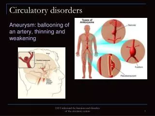
Acute write ventricle insufficiency It is developed by acute stagnation in the big circle of a circulation: • - A swelling of cervical veins; • - Sharp and morbid augmentation of a liver; • - Edemas on the inferior extremities; • - Plus signs of disease which has led acute r\v insufficiency: a pulmonary heart, a thrombembolism of a pulmonary artery.
Acute vascular failure • Syncope - an acute vascular failure with a short-term loss of consciousness that is caused by an acute anemia of a brain.
The basic signs: • Unexpected or instant switching-off of consciousness for some seconds till 1-2 minutes, is rare up to 5 minutes • Before this it can be observed: delicacy, giddiness, a ear noise, emptiness in a head, darkening in opinion of, paleness, a sweating, nausea, a numbness of extremities. • Objectively: paleness, a sweating, a cold snap of extremities, small sphygmus speeded up, arterial pressure is depressed, respiration infrequent, superficial, tendon jerks are conserved. At a long syncope twitching of muscles of the face and extremities can be observed convulsive, salivation strengthens. • After a syncope state, during several hours’ delicacy, a headache is observed.
Acute management at syncope: • Horizontal position with the raised inferior extremities; • To provide access of fresh air (to unbutton clothes to air a room); • To impose with heaters to cover a blanket; • To moisten the face and a breast with cold water; • To pound legs and arms cloth or a brush to yield to smell cotton with spirit of ammonia; • Subcutaneously to introduce 1-2 ml. Cordiaminum, 1мл. 10 % of solution of a coffeine, 2 ml. Camphors; • After appearance of consciousness to give a drink hot tea, coffee.
Collapse- more serious form of a vascular failure which arises owing to disturbance of a regulation of a vascular tonus. Clinic of a collapse. • The collapse arises as complication at pathological states and at serious diseases. • Falloff of arterial pressure (below 90 мм.рт.ст), diastolic and pulse pressure; • Paleness of skin, a sweating, depression of temperature; • Threadlike puls, falling off of veins, a mydriasis, its flaccid reaction to light; • Retardation (but not loss) consciousnesses, a desorientation; • The Oliguria or anuria; • Lungs: respiration vesicular with hard shade.
Acute management at a collapse: • Horizontal position of the patient with the alighted head; • To cover, put a heater to legs; • Massive inhalation of the wetted oxygen; • I\v slowly: - 0,3 - 0,5 ml. 1 % of solution of a phenylephine hydrochloride in 10 ml 0,9 % from tonic solution, or i\v 1 ml. 0,1 % of solution of Noradrenalinum, or i\v 2 mg. Cordiaminum in 20 ml. 40 % of a glucose, Atropinum i\v 0,1 % - 1 ml. (at a collapse owing to conducting новокаинамида); • At absence of effect i\v 200 ml. Polyglucinum with 1 ml 1 % of solution of a phenylephine hydrochloride or 0,5 mg. 0,1 % of Noradrenalinum; • After normalization of arterial pressure, spend hospitalization to chamber of an intensive care laying on a stretcher with drained
PULMONARY THROMBOEMBOLISM • It is occlusion of PA by thrombi which primary appear in veins of great circulation or in right part of the heart.
Clinical classification • Lightning – development of symptoms throughout a minute • Sharp- development of symptoms throughout several hours • Subsharp - development of symptoms throughout several days • Relapsed - at repeatedly of main symptoms • Erased
Clinical classification Behind degree of defeat of a pulmonary vessel: • Deadly - occlusionof 75 % of a pulmonary vascular channel there are more • Massive - defeat is more than 50 % of a pulmonary vascular channel • Sub massive- defeat is less than 50 % of a pulmonary vascular channel • Small or small branches - defeat in суме is less than 25 % of a pulmonary vascular channel
PULMONARY THROMBOEMBOLISM • Thrombi forming anywhere in the venous circulation can embolize in the lung. • In practice more than 90% of clinically significant emboli arise in the deep veins of the legs and thighs and are associated with venous stasis.
PULMONARY THROMBOEMBOLISM • The clinical effects of thromboembolism vary depending on the volume of emboli and on the condition of both the pulmonary and systemic circulations.
PULMONARY THROMBOEMBOLISM The effects of embolism on gas exchange • increased dead space ventilation, • pneumoconstriction, • impaired synthesis of pulmonary surfactant
PULMONARY THROMBOEMBOLISM • The ventilation of unprefused lung adds to the work of breathing and produces tachypnea and a sense of dyspnea. • Some patients manifest asthma-like wheezing. • Impaired surfactant production is a delayed effect that produces edema and atelectasis.
PULMONARY THROMBOEMBOLISM • Massive pulmonary embolism is a well-recognized cause of sudden death, which may be virtually instantaneous or extend over a period of a few minutes. • The major pulmonary arteries are distended with clots that are often coiled or twisted and bear the imprint of venous valves. • The lung parenchyma shows little change except congestion, which presumably comes by way of the bronchial circulation.
Summary • Occlusion of pulmonary arteries, are almost always embolic. • Thrombosis of pulmonary arteries, are rare and occurs only in pulmonary hypertension and pulmonary atherosclerosis. • More than 95 % of pulmonary emboli arise in deep veins of legs. • Frequency of pulmonary embolism correlates with a predisposition to thrombosis in the legs. 30% of the patients with severe burns, trauma or fractures show pulmonary embolism at autopsy.
Summary • Large emboli impact in the major pulmonary artery or astride the bifurcation of the pulmonary artery (saddle embolus). • Emboli may cause instantaneous death. • Emboli may cause cardiovascular collapse
Summary • Hemodynamic compromise is secondary, not only to vascular obstruction, but also to reflex vasoconstriction caused by such agents as thromboxane A2+. • Small emboli may be clinically silent in patients without cardiovascular failure.Emboli may cause transient chest pain and sometimes haemoptysis due to pulmonary hemorrhage. • Pulmonary hemorrhage is characterized by blood in the alveoli, but there is no ischemic necrosis of the pulmonary parenchyma .
Summary • In patients with compromised pulmonary circulation (Eg. cardiac failure), emboli may give rise to infarction. • Middle sized emboli occlude moderate-sized peripheral pulmonary branches. • In rare cases, multiple small emboli may produce chronic cor-pulmonale and eventually pulmonary hypertension and vascular sclerosis.
Pulmonary Thromboembolism • Fat embolism is the result of abrupt pressure changes in the long bones, which rupture thin walled venous sinuses and force marrow fat into them. It embolizes to the lung. In addition, levels of plasma triglycerides, free fatty acids, and lipase rise as part of the stress response. Endothelial damage is caused by fatty acids released from embolized fat and by mediators released during associated blood coagulation. Fat emboli can be recognized by ordinary histopathologic sections as sharply delimited, empty-appearing capillary loops or arterioles, but frozen sections stained for fat are required for confirmation.
Pulmonary Thromboembolism • Amniotic fluid emboli are a rare complication of pregnancy. Infusion of amniotic fluid occurs during tumultuous uterine contractions when the head is in the birth canal. The amniotic fluid is forced through a rupture in the chorion into the maternal veins, precipitating severe dyspnea, tachypnea, and hypotension. Disseminated intravascular coagulation is a common consequence. At autopsy the lungs are hemorrhagic. Squamous cells are lodged in the arterioles. Amniotic debris also contains lipid and mucin, which can be identified with appropriate stains. Reportedly, the clinical diagnosis can be confirmed by the demonstration of squamous cells in blood withdrawn by pulmonary artery catheter.
Pulmonary Thromboembolism • Air embolism can be produced during inspiration if negative intrathoracic pressure draws air into an open vein, an event most likely to happen during a neurosurgical or ear, nose, and throat procedure in which the patient sits upright and the operative wound is above the level of the heart. Air bubbles become trapped in pulmonary arteries and right ventricle where they mechanically impede blood flow. Reactions at the gas-fluid interface trigger blood clotting and the accumulation and activation of neutrophils. Small fibrin and platelet thrombi are found in pulmonary arteries. The physiologic consequences include transient airway constriction and vasoconstriction with great increases in pulmonary vascular resistance and pulmonary artery pressure. With large emboli pulmonary edema, hypoxemia, systemic hypotension and myocardial ischemia are seen. Fatalities have been reported with embolism of 100 ml of air.
Pulmonary Thromboembolism • Foreign-body embolism can result from introduction of foreign material into the veins during medical procedures but is also common among intravenous narcotic users. Particles of insoluble material added as “fillers” to drugs intended for oral use embolizes to the lung and impact in arterioles and small muscular arteries where they cause thrombosis and proliferation of intimal cells. Often they migrate into the perivascular space or interstitium where they give rise to foreign-body granulomas composed of macrophages, multinucleated giant cells, and a few lymphocytes. The process of migration appears to involve the production of granulomatous response in the vascular wall with disintegration of muscle and elastic tissue. In cases where lesions are not numerous, their detection is aided by the use of polarizing filters, since cornstarch and talc, two of the materials commonly used as fillers, are strongly birefringent. When the vascular thrombosis is widespread, pulmonary hypertension results. Lesions may resemble those of primary pulmonary hypertension, particularly in view of the cellular proliferation induced by the foreign material. Extensive interstitial granulomas can produce roentgenographic nodularity and a restrictive ventilatory defect.
Circulatory Disorders • Drugs used are to maintain, preserve or restore circulation • Anticoagulants & antiplatelets (antithrombotics), thrombolytics, antilipemics, peripheral vasodilatiors • Anticoagulants - prevent formation of clots that inhibit circulation • Antiplatelets - prevent platelet aggregation • Thrombolytics (clot busters) - attack/dissolve formed clots • Antilipemics - decrease bld. lipid concentration • Peripheral vasodilators - promote dilation of vessels narrowed by vasospasm
Circulatory Disorders Thrombus Formation • Clot is a Thrombus formed in an arterial or venous vessel • thrombophlebitis - Both inflammation and clots are present • Some thrombus can be superficial but it’s the DVT that’s a concern embolism to lungs.
Circulatory Disorders Thrombus Formation • Arterial formation - begins w/ platelet adhesion to arterial vessel wall Adenosine diphosphate (ADP) released from platelets more platelet aggregation Bld. flow inhibited fibrin, platelets & RBC’s surround clot build up of size structure occludes bld vessels tissue ischemia • The result of Arterial Thrombus is localized tissue injury from lack of perfusion
Circulatory Disorders Thrombus Formation • Venous Formation - Usually from slow bld flow - Can occur rapidly Stagnation of the blood flow initiate the coagulation cascade production of fibrinenmeshes RBC’s & platelets to form the thrombus. Venous thrombus has a long tail that can break off to produce an embolus. These travel to faraway sites then lodge in lung (capillary level) inadequate O2 & CO2 exchange occur (ie. pulmonary embolism & cerebral embolism) • Oral & parenteral anticoagulants (Heparin/Warfarin) primarily act by preventing venous thrombosis • Antiplatelet drugs primarily act by preventing arterial thrombosis
Circulatory Disorders Thrombus Formation • Hemostasis is the normal homeostatic process of blood clotting. • Clotting proteins normally circulate in an inactive state & must be activated to form a fibrin clot. When there is a trigger - inc. bld viscosity from bed rest & stasis - the clotting cascade is activated. • Bld vessel injured platelets adhering to site of injury release of ADP a platelet plug - is ex. of Intrinsic clotting path. • Tissue injury (outside bld vessels) = extrinsic pathway activated
Circulatory Disorders Thrombus Formation • Risk Factors for Deep Vein Thrombophlebitis and Thromboembolism • Three factors increasing risk 1) Stasis of venous flow, 2) damage of the endothelium(inner lining of vein), and 3) hypercoagulability of the blood. • Hx. of thrombophlebitis, abdominal & pelvic surgery, Obesity, neoplasms (lung), CHF, Advanced age, A-fib, vasospasm, Prolonged immobility (bed-rest, long trip spinal cord injury, FX. hip), CVA MI PG, post partum, Estrogen TX (oral contraceptives), IV therapy, trauma, Sepsis, Venous cannulation, Drug abuse, Cigarette smoking Excessive vit E intake Hypercoagulable states (Polycythemia, severe anemias, Dehydration or malnutrition), Antithrombin III deficiency
Circulatory Disorders Anticoagulants • Inhibit clot formation - Do NOT dissolve clots already formed, but prophylactically prevent new clots • Used in clients w/ venous/arterial disorders that put them at inc. risk of clot formation • Venous = DVT & Pulmonary embolism • Arterial = Coronary thrombosis (MI), artificial heart valves, CVA
Circulatory Disorders Heparin • A natural substance in the liver that prevents clot formation. • Primary use is to prevent venous thrombosis that can lead to pulmonary embolism (PE) or stroke • Combines w/ antithrombin III inactivates thrombin and other clotting factors then the conversion of fibrinogen to fibrin doesn’t occur so the clot is prevented • Poorly absorbed through GI mucosa - given SQ & IV • Prolongs clotting time - partial thromboplastin time (PTT) & activated partial thromboplastin time (aPTT) - both bld tests are monitored during therapy
Circulatory Disorders Heparin • Use - DVT, PE, & CVA, Rx of clients w/ heart valve prosthesis, during CV surgery, post op, during hemodialysis * Low doses = prophylactically to prevent DVT * Full doses = treats a thromboembolism & promotes neutralization of activated clotting factors = prevents extension of thrombi & formation of emboli * If started shortly after formation of a thrombus - heparin will also prevent it from developing into an insoluble stable thrombus = reduced tissue damage
Circulatory Disorders Heparin • SE - Decreased platelet count = thrombocytopenia Hemorrhage - give protamine sulfate IV (an anticoagulant antagonist) • DI - Inc. effects w/ ASA, NSAIDs, thrombolytics Dec. effect w/ NTG
Circulatory - LMWH • Low Molecular Weight Heparins (LMWHs) - recently introduced to prevent venous thromboembolism • Binds to Antithrombin III which inhibits the synthesis of factor Xa & formation of thrombin • - enoxaparin (Lovenox) & dalteparin sodium (Fragmin) • - more stable dose, lower risk of bleeding, freq. lab monitoring not required
Circulatory DisordersLMWHs • Use - Prevention of DVT after hip & knee replacement surgery & abd. surgery • Can be administered at home • Administered SQ BID • Available in prefilled syringes w/ attached needles • Usually given in the abdomen • Average Rx is 7 to 14 days • Bleeding less likely to occur • DI - caution client not to take antiplatelet drugs (ASA) during therapy
Circulatory DisordersWarfarin (Coumadin) • Action - Inhibits activity of vit. K required for the activation of clotting factors II, VII, IX, & X. Blocking these factors prevents clot formation • Use - prophylactically to prevent venous thrombosis, A. fib., PE, coronary occlusion, thrombophlebitis • Prolongs clotting time & is monitored by the lab bld. tests prothrombin time (PT) & International normalized ratio (INR) - usually before administering the next dose until therapeutic levels are reached. INR is 1.3 - 2.0 therapeutic levels on coumadin = 2.0 - 3.0
CIRCULATORY DISORDERSWarfarin (Coumadin) • INR is replacing the PT INR more accurate. Need higher levels for prosthetic heart valves, cardiac valvular disease and recurrent emboli. • PT not consistent lab to lab or reagents used. • PT is 1.5 – 2 times the reference value to be therapeutic • Regular monitoring is required for the duration of drug therapy • Warfarin is well absorbed through the G.I. tract. Food decreases.
Circulatory DisordersAntiplatelet Drugs Aspirin, Dipyridamole (Persantine), Ticlopidine (Ticlid) abciximab (ReoPro), tirofiban (Aggrastat) • Action: To prevent thrombosis in the arteries by suppressing platelet aggregation via diff. methods • Use: Prevention of MI/stroke for clients w/ family hx - prevention of a repeat MI, stroke in clients having TIA’s • Persantine & Ticlid = similar to ASA but more expensive • ReoPro & Aggrastat = mainly for acute coronary syndromes. Route = IV
Circulatory DisordersThormbolytics • Thromboembolism - Occlusion of an artery or vein caused by a thrombus or embolus - results in ischemia that causes necrosis of the tissue distal to the obstructed area. - it takes about 1 to 2 weeks for the blood clot to disintegrate by natural fibrinolytic mechanisms - if new thrombus dissolved quicker damage minimized & bld flow restored faster purpose of therapy • Thrombolytics promote fibrinolytic mechanism (convert plasminogen to plasmin & destroys the fibrin in the clot) - administering a thrombolytic drug = clot disintegrates
Circulatory DisordersThrombolytics • Use = Acute MI - w/ in 4 hrs to dissolve clot & unblock artery, so decrease necrosis to myocardium & hospital stay is decreased. • Other uses: Pulmonary embolism, DVT, Noncoronary arterial occlusion • Streptokinase, Urokinase, Tissue plasminogen activator (t-PA), anisoylated plasminogen streptokinase activator complex (APSAC) • Streptokinase & Urokinase are enzymes that act to convert plasminogen to plasmin • t-PA and APSAC activate plasminogen by acting specifically on clot.
Circulatory DisordersAntilipemics • Used to Lower bld. lipid levels • Cholesterol, triglycerides & phospholipids transported in the body bound to protein in various amounts - chylomicrons, very low-density lipoproteins (VLDL), low-density lipoproteins (LDL), high-density lipoproteins (HDL) - more protein & less lipid (removes chol. from bld. stream & deliver it to the liver) • VLDL & LDL contribute to atheroslerotic plaque in bld vessels - composed of mainly cholesterol & triglycerides