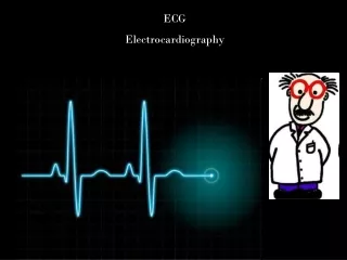
Electrocardiography
Electrocardiography. Prof. K. Sivapalan. Principle of Electrocardiogram. Trunk as volume conductor. Positively charged and negatively charged areas create electrical field. The field changes during spread of impulse. Depolarization towards – upward deflection & away – downward deflection.

Electrocardiography
E N D
Presentation Transcript
Electrocardiography Prof. K. Sivapalan
Principle of Electrocardiogram. • Trunk as volume conductor. • Positively charged and negatively charged areas create electrical field. • The field changes during spread of impulse. • Depolarization towards – upward deflection & away – downward deflection. ECG
Einthovens triangle. • When the source of the electrical field is at the centre of an equilateral triangle, the sum of electrical potential at the three angles is zero. • Since the heart is roughly at the centre of the triangle and the source moves, 5000 ohm resistance is added to the three leads to get zero potential. ECG
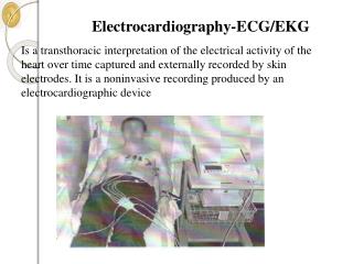
Electrocardiogram. • Electrocardiogram is the record of the changes in the electrical field in the body produced by the electrical activity of the heart. • It represents the sequence and strength of the electrical events in the heart. ECG
Recording ECG. Coronal plane: • Limb leads [Bipolar leads]. • Augmented limb leads.[unipolar] Transverse plane: • Chest [Unipolar] leads V1 to V6 across the chest. Lead I aVR aVL Lead III Lead II aVF ECG
Typical Electrocardiogram. • Paper speed- 25 mm/sec. • Vertically, 1 mV = 10 mm. • P wave – first wave. • Q wave – first down. • R wave – first up in QRS. • S wave – down after R. • T wave – after QRS. • U wave – after T. • PR interval – 0.18 sec. • QRS duration – 0.08 sec. ECG
Information from ECG. • Rate. • Rhythm • Pattern of spread of impulse and integrity of conducting system. • Size [thickness] of myocardium. • Electrolyte imbalance. • Damage to myocardium. ECG
Examples of abnormal ECG. SA node block ECG
Examples of abnormal ECG. Normal. K+ – 4 – 5.5 meq / L K+ – 7 meq / L. K+ – 8.5 meq / L K+ – 3.5 meq /L. K+ – 2.5 meq / L. ECG