Neural Zones
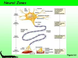
Neural Zones. Figure 5.2. How Neurons connect. The Synapse. A functional connection between surfaces Signal transmission zone Synapse – synaptic cleft, presynaptic cell, and postsynaptic cell Synaptic cleft – space in between the presynaptic and postsynaptic cell

Neural Zones
E N D
Presentation Transcript
Neural Zones Figure 5.2
The Synapse • A functional connection between surfaces • Signal transmission zone • Synapse– synaptic cleft, presynaptic cell, and postsynaptic cell • Synaptic cleft – space in between the presynaptic and postsynaptic cell • Postsynaptic cell – neurons, muscles, and endocrine glands • Neuromuscular junction – synapse between a motor neuron and a muscle
The Synapse • Axon terminal: found in motor neurons • Axon varicosities: ie swellings. Arranged like beads on a string and contain neurotransmitter containing vesicles • En passant synapse: CNS. Consists of a swelling along the axon • Spine synapse: presynaptic cell connects with a dendritic spine on the dendrite of the postsynaptic cell * * * *
The Synapse • Axodentritic: between axon terminal of one neuron and the dendrite of another • Axosomatic: between the axon terminal of one neuron and the cell body of another • Dendrodendritic: between dendrites of neurons (often are electricla synapses) • Axoaxonic: between an axon terminal of a presynatpic neuron and the axon of a postsynaptic neuron. * * * *
Diversity of Signal Conduction • So far: • Electrotonic • Action potentials • Saltatory conduction • Chemical and electrical synapses
Diversity of Synaptic Transmission Figure 5.26
Electrical synapses • cells connect via gap junctions • membranes are separated by 2 nm • gap junctions link the cytosol of two cells • provide a passageway for movement of very • small molecules and ions between the cells • gap junction channels have a large conductance • NO synaptic delay (current spread from cell to cell is instantaneous) - important in some reflexes • chemical synapses do have a significant delay ie slow • commonly found in other cell types as well i.e. glia • can be modulated by intracellular Ca2+ , pH, membrane voltage, calmodulin • clusters of proteins that span the gap such that ions and small molecules can pass directly from one cell to another
More about electrical synapses • cells connect via gap junctions • - made up of 6 protein subunits arranged around a central pore, made up of the connexin protein • - the two sides come together to make a complete unit of 12 proteins around the central pore
Chemical Synapse Diversity • Vary in structure and location Figure 5.27
Chemical Synapse • most common type of synapse • electrical signal in the presynaptic cell is communicated to the postsynaptic cell by a chemical (the neurotransmitter) • separation between presynaptic and postsynaptic membranes is about 20 to 30 nm • a chemical transmitter is released and diffuses to bind to receptors on postsynaptic side • bind leads (directly or indirectly) to changes in the postsynaptic membrane potential (usually by opening or closing transmitter sensitive ion channels) • the response of the neurotransmitter receptor can depolarizes (excitatory postsynaptic potential; epsp) or hyperpolarizes (inhibitory postsynaptic potential; ipsp) the post-synaptic cell and changes its activity • significant delay in signal (1 msec) but far more flexible than electrical synapse
More about chemical Synapses • Some types of chemical synapse include • Excitatory - excite (depolarize the postsynaptic cell • Inhibitory - inhibit (hyperpolarize the postsynaptic cell) • Modulatory - modulates the postsynaptic cells response to other synapses
General sequence of events * * * * * * *
General sequence of events • 1. Nerve impulse arrives at presynaptic terminal • 2. Depolarization causes voltage-gated Ca 2+ channels to open- increases Ca 2+ influx, get a transient elevation of internal Ca 2+ ~100 mM • 3. Vesicle exocytosis- increase in Ca 2+ induces fusion of synaptic vesicles to membrane- vesicles contain neurotransmitters • 4. Vesicle fusion to membrane releases stored neurotransmitter • 5. Transmitter diffuses across cleft to postsynaptic side • 6. Neurotransmitters bind to receptor either:i) ligand-gated ion channel or ii) receptors linked to 2nd messenger systems • 7. Binding results in a conductance change - channels open or close or - binding results in modulation of postsynaptic side • Cont…….
General sequence of events • 8. Postsynaptic response - change in membrane potential (e.g. muscle contraction in the case of a motorneuron at a neuromuscular junction) • 9. Neurotransmitter is removed from the cleft by two mechanismsi) transmitter is destroyed by an enzyme such as acetylcholine esteraseii) transmitter is taken back up into the presynaptic cell and recyclede.g. - acetylcholine esterase, breaks down acetylcholine in cleft, choline is recycled back into the presynaptic terminal
Neurotransmitters • Characteristics • Synthesized in neurons • Released at the presynaptic cell following depolarization • Bind to a postsynaptic receptor and causes an effect
Neurotransmitters, Cont. • More than 50 known substances • Categories • Amino acids • Neuropeptides • Biogenic amines • Acetylcholine • Miscellaneous ….. • Neurons can synthesize many kinds of neurotransmitters
Signal Strength • Influenced by neurotransmitter amount and receptor activity • Neurotransmitter amount: Rate of release vs. rate of removal • Release: due to frequency of APs • Removal • Passive diffusion out of synapse • Degradation by synaptic enzymes • Uptake by surrounding cells • Receptor activity: density of receptors on postsynaptic cell
Graded Potentials via Neurotransmitters • Vary in magnitude depending on the strength of the stimulus • e.g., more neurotransmitter more ion channels will open • Can depolarize (Na+ and Ca2+ channels) or hyperpolarize (K+ and Cl- channels) the cell
Graded Potentials Figure 5.4
Graded Potentials Travel Short Distances Figure 5.6
Neurotransmitter Receptor Function • Ionotropic • Ligand-gated ion channels • Fast • e.g., nicotinic ACh • Metabotropic • Channel changes shape • Signal transmitted via secondary messenger • Ultimately sends signal to an ion channel • Slow • Long-term changes Figure 5.28
Second Messenger again • When activated by a ligand the catalytic domain starts a phosphorylation cascade • Named based on the reaction catalyzed
Neurotransmitter receptors • Different types of neurotransmitter receptors • Functional Type Ligand Ion Channel • Excitatory Receptors Acetylcholine Na+/K+ • Glutamate Na+/K+; Ca2+ • Glutamate Na+/K+ • Serotonin Na+/K+ • Inhibitory Receptors Aminobutyric acid, GABA Cl- • Glycine Cl-
Amount of Neurotransmitter • Influenced by AP frequency which influences Ca2+ concentration • Control of [Ca2+] • Open voltage-gated Ca2+ channels [Ca2+] • Binding with intracellular buffers [Ca2+] • Ca2+ ATPases [Ca2+] • High AP frequency influx is greater than removal high [Ca2+] many synaptic vesicles release their contents high [neurotransmitter]
Removal of Neurotransmitter • broken down by enzyme • - acetylcholine esterase breaks down acetylcholine in the synaptic cleft • - many nerve gases and insecticides work by blocking acetylcholine esterase – Yikes! • - prolongs synaptic communication • b) recycled by uptake • - most neurotransmitters are removed by Na+/neurotransmitter symporters • - due to a specific neurotransmitter transporter • - recycled by uptake into presynaptic terminal or other cells (glial cells will take up neurotransmitters) • c) diffusion: simple diffusion away from site
Neurotransmitters - stages • Synthesis • - all small chemical neurotransmitters are made in the nerve terminal • - responsible for fast synaptic signalling • - synthetic enzymes + precursors transported into nerve terminal • - subject to feedback inhibition (from recycled neurotransmitters • - can be stimulated to increase activity (via Ca2+ stimulated phosphorylation) • 2.Packaging into vesicles • - neurotransmitters packaged into vesicles • - packaged in small "classical" vesicles • - involves a pump powered by a pH gradient between outside and inside of vesicle • - pump blocked by drugs and these block neurotransmitter release
Presynaptic vesicles • Two groups i) low molecular weight, non-peptide e.g. acetylcholine, glycine, glutamate • ii) neuropeptide (over 40 identified so far and counting…..)
Presynaptic vesicles • There are 2 types of secretory vesicles • We will only talk about small chemical synaptic vesicles • Neuropeptides are made and packaged in the cell body and transported to synapse) • Small chemical neurotransmitter vesicles • responsible for fast synaptic signaling • store non-peptide neurotransmitters, e.g. acetylcholine, glycine, glutamate • enough vesicles in the typical nerve terminal to transmit a few thousand impulses • exocytosis only occurs after an increase of internal Ca 2+ (due to depolarization) and at active zones (regions in the presynaptic membrane adjacent to the cleft)
Presynaptic vesicles * * * * * * * *
Vesicle Exocytosis • A group of 6 to 7 proteins work together to respond to Ca 2+ influx and regulate vesicle fusion • after exocytosis the synaptic vesicle membranes are reinternalized by endocytosis and reused (reloaded with neurotransmitter by a transmitter transporter system) • vesicles are also transported from the cell body to the nerve terminal- transmitter is synthesized in the terminal and loaded into the vesicles- enzymes and substrates necessary are present in the terminal- i.e. acetylcholine, acetyl-CoA + choline used by choline acetyltransferase
Vesicle Exocytosis • non-peptide transmitters • exocytosis only occurs after an increase of internal Ca 2+ (due to depolarization) • at active zones (regions in the presynaptic membrane adjacent to the synaptic cleft) • peptide-transmitters (same as for non-peptide transmitters except:) • exocytosis is NOT restricted to active zones • exocytosis is triggered by trains of action potentials
SNARE hypothesis The SNARE Hypothesis for Transport Vesicle Targeting and Fusion SNARE is an acronym for SNAP receptor (SNAP stands for soluble N-ethylmaleimide-sensitive factor attachment proteins). SNARES are involved in the mediation of protein transport between various plant organelles by small membrane vesicles. Two families: i) V-SNARE - vesicle membrane proteinsii) T-SNARE - target membrane proteins
SNARE hypothesis • Vesicle docking occurs between the V-SNARE and T-SNARE proteins • The combined proteins act as a receptor for an ATPase that utilizes ATP to generate the "docked" form • One of the proteins is a Ca2+ sensor such that when Ca2+ enters the synapse the vesicle fuses with the plasma membrane and releases its contents • The membrane and proteins are then recycled through endocytosis (clatharin coat and dynamin etc.) and reused.
Acetylcholine • Primary neurotransmitter at the vertebrate neuromuscular junction Figure 5.17
Synaptic Plasticity • Change in synaptic function in response to patterns of use • Synaptic facilitation – APs neurotransmitter release • Synaptic depression – APs neurotransmitter release • Post-tetanic potentiation (PTP) – after a train of high frequency APs neurotransmitter release Figure 5.32
Postsynaptic Cells • Have specific receptors for specific neurotransmitters • e.g., Nicotinic ACh receptors
Diversity of Signal Conduction • So far: • Electrotonic • Action potentials • Saltatory conduction • Chemical and electrical synapses • Also: • Shape and speed of action potential • Due to diversity of Na+ and K+ channels
Ion Channel Isoforms • Multiple isoforms • Encoded by many genes • Variants of the same protein • Voltage-gated K+ channels are highly diverse (18 genes encode for 50 isoforms in mammals) • Na+ channels are less diverse (11 isoforms in mammals) Table 5.2
Channel Density • Higher density of voltage-gated Na+ channels • Lower threshold • Shorter relative refractory period
Voltage-Gated Ca2+ Channels • Open at the same time or instead of voltage-gated Na+ channels • Ca2+ enters the cell causing a depolarization • Ca2+ influx is slower and more sustained • Slower rate of APs due to a longer refractory period • Critical to the functioning of cardiac muscle