Download
1 / 49
500 likes | 692 Vues
Cytolytic Cell Effector Mechanisms. Shabaana Khader, PhD Children’s Hospital Of Pittsburgh 4/6/2009 Shabaana.Khader@chp.edu. Recognition – T cells. T cells recognize antigen via a heterodimeric receptor [TCR] that is clonally distributed

E N D
Cytolytic Cell Effector Mechanisms Shabaana Khader, PhD Children’s Hospital Of Pittsburgh 4/6/2009 Shabaana.Khader@chp.edu
Recognition – T cells • T cells recognize antigen via a heterodimeric receptor [TCR] that is clonally distributed • Heterodimeric TCR is either ab or gd pairing of chains • Antigen is recognized in the context of MHC molecules • CD8 [homo- or heterodimer] binds MHC Class I and serves as a co-receptor • Cytolytic cells are primarily CD8+, but some CD4+ T cells are also cytolytic
A. Activation of naïve, precursor CTLs [pCTLs] in CTLs • To participate in an adaptive immune response, naïve precursor T cells in the periphery must be induced to proliferate and differentiate in response to antigen. • The exposure to antigen occurs in secondary lymphoid organs. • The process requires several days, at which time the terminally differentiated T cells, called armed effector cells, migrate into the periphery and have activity against target cells expressing appropriate antigens. • They differ in several ways from pCTLs, and the changes in these cells aid them in responding rapidly and efficiently when encountering their target cells.
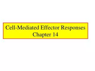
IFNg N CCL19/CCL21 Virus N Migratory DC T Stromal cell CXCR5 CXCL13 T IFNg CCR7 T HEV T T T CCR7 T CCL21 Stromal cell LUNG DRAINING LYMPH NODE DC Chemokines
1. CTL Stimulation Pathways • Role of APCs, secondary lymphoid tissues, T helper cells, cytokines • naïve T cells migrate to secondary lymphoid organs where they encounter antigen presenting cells, e.g. DCs, macrophages and B cells • APCs provide survival signals to naïve T cells, and present antigen in the context of MHC • Activation of naïve T cells requires specific antigen and co-stimulatory signals, TCR/CD28 • Clonal expansion precedes differentiation to effector function • Clonal expansion of CD8+ T cells can be driven by cytokine production by CD4+ T cells, or in some cases directly by autocrine production of cytokines • In either case, IL2 is the primary cytokine driving T cell proliferation and differentiation
1. CTL Stimulation Pathways • Direct priming with endogenous antigens vs cross-priming • CD8+ T cells are generally stimulated by endogenous antigen, expressed in the context of MHC Class I antigens • In some cases, exogenous antigens have been found to be presented in the context of MHC Class I-Use of transgenic mice with specificity for OVA (OT-1) has shown that cross-priming can occur in vivo • Eg/ In vivo DCs can process exogenous antigen from the endocytic to the cytosolic compartment for MHC Class I presentation • Thought to be an important process which facilitates T cell priming to antigens present in non-lymphoid organs (eg. Epithelial cells) which are not normally surveyed by recirculating, naive T cells
Lymphoid organs 1.TCR Signal 2.Strong costimulatory signal Site of Infection TCR signal
In the absence of strong costimulation, CD8 T cells require CD4 T cell help for differentiation of CTLS
CTL differentiation Tc1, Tc2 and Tc17 cells • Similar to CD4+ T cells, clones of CD8+ T cells can be differentiated based upon profiles of cytokine production • IL2 + IL4 in primary cultures of CD8+ T cells induces development of clones secreting IL4 and IL5, termed Tc2 cells • IL2 + IFNg in primary cultures of CD8+ T cells induces development of clones secreting IFNg, termed Tc1 cells (Dutton lab,1994) • IL-2 + IL-1β + IL-6 + and TGF-β induces development of clones secreting IL-17, termed as Tc17 (Hamada et al, Dutton Lab 2009) • Tc1 and Tc2 retain their patterns of cytokine production in vivo: Tc17 less understood
CTL differentiation • Phenotypic alterations • High affinity receptor for IL2 [CD122/CD132/CD25] • Loss of expression of CD62L • Increased expression of CD11a/CD18 [LFA-1] • Increased expression of CD2 • Induction of expression of CD29/CD49d [VLA-4] • Some activated T cells express MHC Class II
Activation of T cells changes the expression of several cell surface molecules CD8 CD8+ High affinity IL2r – CD122/CD132/CD25 MHC Class II – some T cells
CCL19/CCL21 Migratory DC T Stromal cell CXCR5 CXCL13 T IFNg CCR7 T HEV T T T CCR7 T CCL21 Stromal cell LUNG DRAINING LYMPH NODE DC Chemokines
B. The Process Used by Cytolytic Effector Cells • First step is conjugation which is dependent upon adhesion molecules, particularly CD11a/CD18 [LFA-1] and CD2 (on the T cell) binding CD54 and CD58, respectively on the target cell • Second step involves recognition and activation of the lytic machinery of effector cells via one of several signaling pathways. Many observations also indicate a role for LFA-1 and CD2 in activation. • Third step is the delivery of the lethal hit – granule exocytosis, death receptor mediated pathways • These three steps occur rapidly within the context of a short-lived immunologic synapse between CTLs or NK cells and their target cells; • Recycling of the effector cell and delivery of the lethal hit to additional target cells
Lytic granules MTOC-MICROTUBULE ORGANIZING CENTER)
Delivery of Granules to the Immunologic Synapse • On contact with the target cell, the MTOC polarizes towards the target cell • Granules move along microtubules towards the polarized MTOC • Centrosome moves to and contacts plasma membrane • Actin and IQGAP1 are cleared from the synapse • Granules delivered directly to the plasma membrane Stinchcombe, J, et al. 2006. Nature 443:462
Steps in Granule Movement/Exocytosis Bossi, G. et al., 2002. Immunol. Rev. 189:152
CTLs attach to several different target cells and kill them one by one • Quick-preformed lytic granules • Controlled • Less inflammation • Less tissue damage • The same CTL can kill many target cells in succesion
Components of Lytic Granules Form Pores in “Lethally Hit” Target Cells • Perforin monomers bind to phosphorylcholine on the target cell membrane • Ca2+ dependent • Polymerizes to form a pore • Lipophilic on the outside with a hydrophilic central channel 16nm diameter • Water and salts pass rapidly into the cell • Cell death Granzymes
Granzymes • Granzymes are proteases • Granzyme B cleaves and activates Caspase-3-cystiene protease • Caspase 3 activates a caspase proteolytic cascade and activates Caspase-activated deoxyribonuclease(CAD) by cleaving an inhibitory protein (ICAD) that binds to and inactivates CAD. • CAD degrades the DNA • Cells undergo programmed cell death and are rapidly ingested by phagocytes • Phagocytes detect a change in the cell membrane eg. Phosphatidylserine normally found in inner leaflet of the membrane can be detected in the outer leaflet.
Specificity of Granzymes [Grz] GranzymeCategorySpecificityChrmsml. location GrzA* trypsin arg/lys 5q11-12 [h] GrzB** aspase aspartic acid 14q11.2 [h] GrzC ? ? GrzD-G*** GrzH chymase phenylalanine 14q11.2 [h] GrzK trypsin arg/lys 5q11-12 [h] GrzM elastase meth/leu/isoleu 19p13.3 *Caspase-independent induction of apoptosis, slow via induction of single-strand nicks in DNA and prevention repair **Caspase-dependent induction of apoptosis, rapid ***Mice only
Given the nature of perforin-mediated cytotoxicity, what limits autolysis of effector cells? • Highly polarized secretion of perforin • Takes place at the point of contact
Given the nature of perforin-mediated cytotoxicity, what limits autolysis of effector cells? • Cathepsin B is contained in granules of NK cells and CTLs • Cathepsin B is translocated to the cell surface when granule membranes fuse with the plasma membrane • Cathepsin B cleaves perforin at the killer cell surface limiting pore formation • Proposed as a means of protection of the effector cell during target cell destruction
Consequences of Cytolysin Deficiency: Knock Out Mice • Cytolytic cells from perforin-/- mice are deficient in lytic activity in vitro • Perforin-/- mice have increased susceptibility to non-cytopathic and cytopathic viruses; higher titers of Herpes and MCMV • Perforin-/- mice have an increased incidence in lymphomas
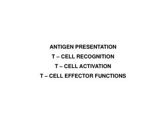
Consequences of Genetic Defects Related to Perforin or Granule Exocytosis • Individuals with perforin mutations – lethal, inherited disease, familial haemophagocytic lymphohistiocytosis [FHL] – believed to be the consequence of a failure to down-regulate an immune response following a viral infection for which protection is strictly perforin-dependent, suggests perforin is important for immune regulation • Deletion of Rab27a, a small GTP-binding protein, results in a lack of ability to exocytose granules [Griscelli Syndrome/Ashen mouse] • Mutation in LYST/CHS1 results in defects in membrane fusion of lysosome/granules which results in the formation of a giant granule which is not exocytosed [Chediak-Higashi Syndrome/Beige mouse] • Mutation in the Rab geranylgeranyl transferase [RGGT] gene results in a failure to translocate granule to the site of the synapse [Hermansky-Pudlak Syndrome/Gunmetal mouse]
Steps in Granule Movement/Exocytosis Reduced NK killing, may be due to less actin accumulation at synapse Granule fusion with membrane Granule motility Granule motility Granule movement from microtubule to synapse Bossi, G. et al., 2002. Immunol. Rev. 189:152
Consequences of Granzyme Deficiency: Knock Out Mice • By comparison to perforin, relatively poorly characterized for in vivo significance • Considerable functional redundancy among granzymes • GrzA-/- mice are basically normal in lytic function and resistance to virus • GrzB-/- mice reduced capacity to induce, and delayed induction of, apoptosis • GrzA/B-/- reduced lytic capacity and increased susceptibility to infection with ectromelia virus
Voskoboinik et al. Nature Reviews Immunology6, 940–952 (December 2006) | doi:10.1038/nri1983
Elimination of intracellular pathogens can be mediated by a novel protein, granulysin, which is associated with granules
Granulysin-mediated Induction of Apoptosis 1) Binds to tumor cell surface on the basis of charge 2) Sphingomyelinase [SM] is activated, resulting in increased ceramide concentration [fast process] 3) Binding results in an increase in intracellular calcium and decrease in intracellular potassium 4) Mitochondria are damaged resulting in release of cytochrome C and apoptosis inducing factor [AIF] [slow process] 5) Activation of caspases and endonucleases resulting in apoptosis Current Opinion in Immunology 2003, 15:560–565 www.current-opinion.com
Granulysin Directly Kills a Variety of Microorganisms Stenger, S., et al. Science 282:121, 1998
Biological Significance and Consequences of Granulysin Deficiency • Granulysin homolog/ortholog has not been found in mice • Potential disease associations with polymorphisms or differences in granulysin as yet unidentified • mRNA in urine may be a very good biomarker for acute graft rejection • siRNA depletion of granulysin resulted in loss of CTL ability to lyse C. neoformans • Required for CTL lysis of M. tuberculosis infected macrophages • Tumor bearing individuals had comparable levels of perforin relative to normals, but remarkably lower levels of granulysin – suggests levels of granulysin have some prognostic value for progression of cancer
Binding of Fas ligand to Fas initiates the process of apoptosis
IFNg N CCL19/CCL21 Virus N Migratory DC T Stromal cell CXCR5 CXCL13 T IFNg CCR7 T HEV T T T CCR7 T CCL21 Stromal cell LUNG DRAINING LYMPH NODE DC Chemokines
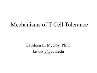
. . . . NK NK NK NK NK NK Cells • Preformed Cytoplasmic granules • No requirement for antigen • Activated by interferons and IL-12 • Can kill target cells by CTL • Can also produce IFNg and • control infection Periphery Stimulus: Hr/Days Cytokines: IFNg GM-CSF TNFa IL2 IL12 IL15 IL21 IL23 IL27 IFNa, -b Proliferation Enhanced CTX, with broader specificity
Recognition – NK cells • There is no evidence supporting clonally restricted recognition molecules expressed by NK cells, nor for recombinatorial events being important for development of an NK cell repertoire • NK cells recognize MHC determinants, but these structures, nor peptides expressed by MHC, are target antigens for activation of NK lytic function • Some NK cells express CD8 homodimers, but it is unclear whether binding to MHC Class I affects activation • NK cell recognition of targets involves a balance between inhibitory signals and activation signals • Receptor:ligand pairs providing inhibitory signals are fairly well defined • Receptor:ligand pairs providing activation signals are rapidly being defined
NK cell activating receptors • Loss of the inhibitory signal does not, in and of itself, provide signals to kill target cells • Some receptors able to activate NK cells to kill target cells have been defined – NKG2D, Ly49D, Ly49H, NKp30, NKp44, NKp46, CD161A • Some activating receptors are members of the C-type lectin [e.g. NKG2D] and IgSF [NKp30] superfamilies • IgSF members often referred to as KARs • Associated with an adaptor molecule [e.g. DAP10] containing an ITAM. Associate via a charged residue in the TM domain • Some ligands for activating receptors have been defined, e.g. RAE-1 for NKG2D
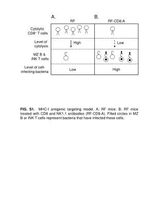
Possible mechanism by which NK cells distinguish infected from UI cells
IFNg N CCL19/CCL21 Virus N Migratory DC T Stromal cell CXCR5 CXCL13 T IFNg CCR7 T HEV T T T CCR7 T CCL21 Stromal cell LUNG DRAINING LYMPH NODE DC Chemokines
Suggested Reading • Janeway Text-Sections 8-12 to 8-25 • Strenger, S et al ,An antimicrobial activity of cytolytic T cells medaited by granulysin. Science 282:121-125(1998) • Trapani et al. The complex issue of regulating perforin expression. Trends in Immunology. 28(6) 243-245 • Yokoyama et al. The dynamic life of natural killer cells. Annu. Rev Immunol. 2004. 22. 405-429 • Clayberger et al. Granulysin. Current. Opinion in Immunol. 2003 15: 560-565