Download
1 / 56
560 likes | 676 Vues
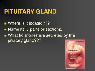
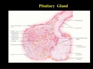
PHYSIOPATHOLOGY OF PITUITARY GLAND, ADRENALS AND GONADS. Hormones of the hypothalamus. Pituitary diseases. Pituitary diseases may be caused by: lesions in the pituitary gland In the anterior or posterior lobe lesions in the hypothalamus Anterior pituitary hyperfunction

E N D
Pituitary diseases • Pituitarydiseasesmay be causedby: • lesionsin the pituitarygland • In the anteriororposteriorlobe • lesionsin the hypothalamus • Anteriorpituitaryhyperfunction • the prolactinomadue to tumor releasingprolactin • acromegaly/gigantismdue to somatotropic adenoma • Cushing’sdiseasedue to corticotropic adenoma • pituitaryhyperthyroidismdue to thyreotropic adenoma • disturbances of reproductivefunctiondue to gonadotropictumors
Prolactinoma • iscomposed of lactotrophiccells • stimulates the production of milk, • causinggalactorrhea • interferes with the normallypulsatilesecretion of luteinizinghormone (LH) and follicle-stimulatinghormone (FSH) • causingamenorrhea, anovulation, infertility, and loss of libido • in men itcauseserectiledysfunction, loss of libido, infertility, and gynecomastia
Causes of Hyperprolactinemia Prolactinoma Physiologicstates Pregnancy, nursing, nipplestimulation Stress Drugs Dopamine receptor antagonists Dopamine-depletingagents Hypothalamic and pituitarystalklesions Craniopharyngioma, surgery, headinjury, irradiation Hypothyroidism Renalfailure Cirrhosis
Somatotropic adenoma • tumor secretesgrowthhormone • gigantism - tumorsin chldchoodfoundbefore the closure of epiphysis • acromegaly - tumorsof adulthood
Acromegaly • Acromegaly • acralenlargement due to thickening of the skin, • excessive growth of subcutaneous tissue and bone, • generalized organomegaly, and metabolic changes 1.Enlargement of hands and thickening of fingers, with palmar skin thickening. 2.Enlargement of feet and toes and heel pad. 3.Facial changes (“acromegalic facies”). prominent supraorbital ridges, enlarged nose, protruding jaw, and enlarged tongue. 4.Enlargement of the head.
Acromegaly 5. Cardiomegaly organomegaly and hypertension, congestiveheart failure 6.Musculoskeletal symptoms Bone overgrowth and weakening of muscle is complicated by degenerative joint disease and widespread pain. 7.Neuropathy Carpal tunnel syndrome and other peripheral nerve compression 8.Metabolic changes glucose intolerance, diabetes
Causes of hypopituitarism • Invasive tumor (e.g., brain tumor of hypothalamus, craniopharyngioma, pituitary adenoma, meningioma, chordomas, metastases) • Iatrogenic (e.g., surgery, radiationtherapy, hormonaltherapy) • Infarction (e.g., Sheehan’spostpartumsyndrome, sicklecell anemia) • Injury (e.g., head trauma) • Infection (e.g., tuberculosis) • Immunedisease (e.g., lymphocytichypophysitis, sarcoidosis) • Inherited (e.g., isolatedgeneticpituitaryhormonedeficiencies, hormone receptor deficiencies) • Idiopathic
Aldosterone- • mineralocorticoid • acts on distaltubules and collectingducts to promoteuptake of Na+ in exchange for K+ or H+. • Cortisol- • glucocorticoid • hasanti-insulin activity, decreasingglucoseuptake in muscle and adiposetissue • enhances the effects of glucagons and epinephrine • Enchancedglycogenolysis and lipolysis • hasanti-inflammatoryeffects • inhibits ADH • Androgens- • androstenedione, dehydroepiandrostenedione (DHEA) dehydroepiandrostenedionesulfate (DHEAS)
Adrenal cortical diseases Hypersecretion of corticosteroids may result from: 1.Hyperplasia of adrenal cortical cells 2.Benign tumors (adenomas) 3.Malignant tumors (carcinomas) 1. Conn’s syndrome – hyperaldosteronism an excess of aldosteroneleads to hypertension and hypokalemia 2.Cushing’s syndrome (hypercortisolism) an excess of glucocorticoids leads to disturbances of the metabolism of carbohydrates, lipidsand proteins 3.Adrenogenital syndrome an excess of androgenic hormones
Cushing’s syndrome 1. Pituitarycorticotropic adenoma (70%) 2. Adrenalcortical adenoma, carcinoma, orhyperplasia (20%) 3. Ectopic ACTH overproduction(10%) - small-celllungcancer, bronchialorgastrointestinalcarcinoidtumors 4. Iatrogenic (administration of glucocorticoids for immunosuppression)
Cushing’s syndrome • Weight gain, moon face, truncal obesity, and accumulation of fat on the back • cortisol-induced hyperglycemia and reactive hyperinsulinemia - fat accumulation • Hyperphagia - increase of appetite • muscle wasting on the extremities and anterior abdominal wall • thin arms and legs and weak abdominal muscles • atrophy of the skeletal muscles caused by increased proteolysis • Hypertension • due to the mineralocorticoid effects of cortisol • retention of sodium and water in the kidneys
Cushing’s syndrome • Glucose intolerance • cortisol has anti-insulin activity and stimulates gluconeogenesis and glycogenolysis in the liver • Skin changes - due to effects of cortisol on collagen synthesis : • skin striae • weakness of the connective tissueand vascular fragility • delayed wound healing • Osteoporosis • Cortisol • inhibits collagen synthesis • accelerates bone breakdown • healing of fractures is delayed • Polycythemia - increased red blood cell count • cortisone stimulates erythropoietin
Cushing’s syndrome • Reproductive changes and virilization • menstrual irregularities (oligomenorrheaor amenorrhea, infertility) • signs of virilization, such as hirsutism, and an increased incidence of acne on the face and the back. • thesechanges are related to the androgenic effects of 17-ketosteroids. • males suffer from impotence and a loss of libido • Depression, sleep disturbances, and memory loss • Patientsinitially euphoric later depressed • Sleep disturbances l insomnia or lethargy • Short-term memory impairement
Hyperaldosteronism Two forms of hyperaldosteronism: • 1.Primary hyperaldosteronism (Conn’s syndrome). • cortical adenomas (75%) • idiopathic • 2.Secondary hyperaldosteronism. • the influence of renin -angiotensin II - aldosterone
In primary hyperaldosteronism excess of aldosterone suppresses the secretion of renin (hyporeninemic hyperaldosteronism). • In secondary hyperaldosteronism both the renin and aldosterone levels are high (hyper-reninemic hyperaldosteronism).
Hyperaldosteronism 1. Hypertension • Headaches • hypertensive retinopathy 2.Hypernatremia Aldosterone leads to a retention of sodium (Na+), leading to initial hypernatremia. Elevation of Na+ in serum leads to a reactive release of atrial natriuretic hormone (ANH) 3.Hypokalemia • excretion of potassium (K+) in urine • Hypokalemia may cause muscle weakness and fatigue. • The ECG shows changes such as flattening of T waves and the appearance of U waves
Hyperaldosteronism 4.Nocturnal polyuria (hypokalemic nephropathy), kidneysresistant to ADH • kidneys cannot retain the water, polyuria inducespolydypsia • Loss of K+ is associated with retention of bicarbonate (HCO3−) -metabolic alkalosis 5.Metabolic alkalosis • hypokalemia, the intracellular K+ isreleased into the blood. • The loss of intracellular K+ is leads tointracellular influx of H+ and Na+. • The loss of H+ causes metabolic alkalosis.
Clinical signs and symptoms • 1.Weakness and easyfatigability • 2.Weight loss and anorexia • 3.Hyperpigmentation of the skin and mucousmembranes • 4.Nausea and vomiting • 5.Abdominal pain • 6.Diarrhea orconstipation • 7.Electrocardiographic changes of hyperkalemia, such as high-peaked T waves and a prolonged PR interval • 8.Cardiac arrhythmia, includingcardiacblock • 9.Salt craving • 10.Orthostatic hypotension and syncope
Diseases of the Adrenal Medulla Tumors: • Neuroblastoma - in childhood • tumors are composed of primitive neuroblastic precursors of adrenal medullary cells • Pheochromocytoma - in adults • the tumors are composed of well-differentiated cells secreting epinephrine or norepinephrine
Pheochromocytoma 1.Heart—tachycardia and stronger myocyte contraction 2.Blood vessels—vasoconstriction, but also vasodilation in some vessels 3.Kidney—renin release 4.Liver—glycogenolysis leading to hyperglycemia 5.Fat tissue—lipolysis 6.Intestine—smooth muscle cell relaxation and loss of peristalsis 7.Skin—sweating with cold and pale extremities due to constriction of peripheral arterioles
Pheochromocytoma 1. Hypertension results from vasoconstriction of arterioles and increased cardiac output -better venous return -positive inotropic effect of catecholamines on the myocardium In about 50% of cases the hypertension issustained 2.Sympathetic effects headache, sweating, palpitations, tachycardia, facial flushing, nausea, vomiting 3.Posturalhypotensive episodes and syncope 4.increasedmetabolic rate fever with sweating, tachycardia, easy fatigability, weight loss 5.Hyperglycemia Catecholamineshave an anti-insulin effect and promote glycogenolysishyperlipidemia -lipolytic effect of catecholamines on peripheral fat stores
The testes are under the control of the hypothalamic GnRH pituitary gonadotropic hormones FSH and LH Gonadotropic hormones stimulate the Sertoli and Leydig cells, which in turn cooperate in supporting spermatogenesis. Sertoli cells function is to support spermatogenesis. produce a polypeptide called inhibin, which inhibits the release of pituitary gonadotropins by negative feedback. Leydig cells the primary source of testosterone and other androgens in the male body. Androgens act locally on Sertoli cells and spermatogenic cells and are also released into the circulation.
TESTOSTERONE • Promotes the growth of the penis, seminiferous tubules, and scrotum • Stimulates formation and maturation of spermatozoa • Stimulates secretory activity of the prostate and the seminal vesicle • Increases erythropoiesis • Promotes muscle growth (protein anabolism) • Promotes bone growth, mineralization and fusion of the epiphyseal plates • Stimulates laryngeal growth • Causes hair loss in the male • Stimulates libido and aggressive behavior • Stimulates renal retention of electrolytes • Reduces the concentration of HDL in blood • Influences fat distribution
DECREASED RELEASE OF ANDROGENS • Causes • Lack of GnRH or nonpulsatile secretion of GnRH • Damage to the hypothalamus • Tumor • Radiation • Abnormal perfusion • Genetic defect • Psychological or physical stress • Persistently high concentrations of GnRH down-regulation of the receptors decreased gonadotropin release
DECREASED RELEASE OF ANDROGENS • Inhibition of gonadotropin release • Prolactin • Damage to the pituitary • Trauma • Infarct • Autoimmune disease • Tumor • Hyperplasia • Damage to the testes • Genetic defect • Severe systemic disease • Enzyme defects in hormone synthesis • Defect of testosterone receptors
DECREASED RELEASE OF ANDROGENS Effects: • In male fetus – absent sexual differentiation • In juveniles • Failure of the voice break • Absence of adult body hair • Delayed bone growth • Delayed epiphyseal fusion excess longitudinal growth of the limbs • In adults • Infertility • Reduced libido and aggressiveness • Reduces muscle and bone mass • Increased hematocrit
ANDROGEN EXCESS Causes: • Enzyme defects in steroid hormone synthesis • Testosterone producing tumor • Iatrogenic androgen supply Effects: • Male sex differentiation • Hair growth in females • Increase in erythropoiesis • Increased muscle and bone mass • Increased libido and aggressiveness • Amenorrhea in females • Decreased GhRH and gonadotropin infertility in males and females
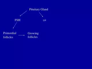
GONADOTROPIC HORMONES IN FEMALES • LH and FSH released from the pituitary anterior lobe in pulsatile manner (every 60 to 90 minutes for 1 min.) • Stimulated by pulsatile release of GnRH from the hypothalamus Effects of FSH: • Promotes the maturation of the follicles • Stimulates estrogen production in the granulosa cells of the follicles Effect of LH: • Stimulates follicle rupture and ovum discharge • Stimulates corpus luteum formation • Stimulates progesterone synthesis
Duringfollicularphase: • Estrogens (estrone, estradiol, estriol) stimulaterelease of gonadotropins – positive feedback • Duringlutealphase: • Progestogens (progesterone and analogs) and estrogensinhibitfurtherrelease of gonadotropins (negative feedback) • Granulosacellsproduceactivin (promotes gonadotropin release) and inhibin (suppressesit).
Hypothalamic-pituitary-ovarian axis. Gonadotropin-releasing hormone (GnRH) stimulates the pituitary, which secretes the follicle-stimulating hormone (FSH) luteinizing hormone (LH). FSH and LH stimulate granulosa and theca cells to produce estrogens, progestins, androgens, inhibins, and activins, which provide negative or positive feedback to the hypothalamus or the pituitary. Inhibin inhibits the pituitary activins activate it.
Hormonal changes during the menstrual cycle. The surge of luteinizing hormone (LH) on day 13 leads to ovulation, a point that divides the proliferative from the secretory phase. Estrogen is the prevalent ovarian hormone during the proliferative (follicular) phase. Progesterone predominates in the secretory phase. FSH, follicle stimulating hormone.
ESTROGENSPHYSIOLOGICAL ACTION • Promote the development of the female sex characteristic • The secondary sexual characteristics • Development of the mammary glands • Female fat distribution • Promote bone growth and maturation and accelerate epiphyseal fusion • Influence the psychological development of women • Increase the coagulability of blood • Raise electrolyte retention in the kidneys • Increase mineralization of the bones • Hydroxylation of vitamin D3 • Inhibition of parathyroid hormone (PTH)
PROGESTERONEPHYSIOLOGICAL ACTION • Promotes the maturation and secretory activity of the uterine mucosa • Decreases the contractility of the uterine muscle • Raise the body’s metabolism and temperature • Trigger hyperventilation • Reduce sensitivity to insulin in the periphery • Moderate glucocorticoid and antimineralocorticoid (natriuretic) actions • Lower the production of cholesterol and the plasma concentration of HDL and LDL.
FEMALE SEX HORMONES EXCESS Causes: • Exogenous supply (contraceptive pills) • Tumors producing sex hormones • Folliculoma • Thecoma
DEFICIENCY OF GONADOTROPINS Causes: • Decreased GnRH release • Malnutrition • Severe systemic disease • High performance sport • Influence of neurotransmitters: norepinephrine, dopamine, serotonin, endorphins • Persistently high concentrations of GnRH down-regulation of GnRH receptor