O-Glycosylation
650 likes | 666 Vues
This lecture delves into the intricate world of O-linked glycosylation, discussing structures, functions, and regulatory mechanisms of O-Glycosylation pathways. Explore mucins, proteoglycans, and distinctive glycan linkages in different proteins. Dive into the roles of O-Fuc and O-Glc in modulating signaling, with a focus on diseases related to disrupted O-Glycosylation.

O-Glycosylation
E N D
Presentation Transcript
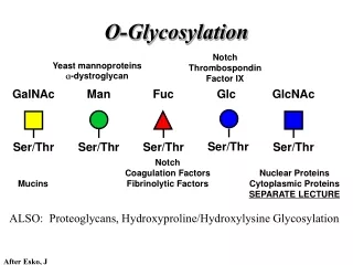
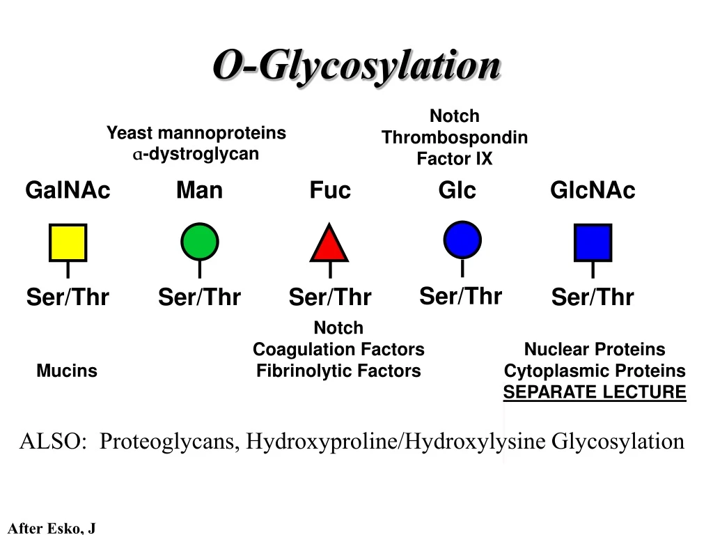
Glc GlcNAc GalNAc Man Fuc Ser/Thr Ser/Thr Ser/Thr Ser/Thr Ser/Thr O-Glycosylation Notch Thrombospondin Factor IX Yeast mannoproteins a-dystroglycan Notch Coagulation Factors Fibrinolytic Factors Nuclear Proteins Cytoplasmic Proteins SEPARATE LECTURE Mucins ALSO: Proteoglycans, Hydroxyproline/Hydroxylysine Glycosylation After Esko, J
O-glycosidic linkage is sensitive to alkali (regardless of stereochemistry) b-elimination O-Glycosidic Linkage GalNAc GalNAc a a Ser After Esko, J
Glycan synthesis in a cellular context Most O-Glycosylated proteins are synthesized in the secretory pathway
Glc GlcNAc GalNAc Man Fuc Ser/Thr Ser/Thr Ser/Thr Ser/Thr Ser/Thr O-Glycosylation
a3 b4 a3 b3 b4 b3 b4 a3 b3 b6 Ser/Thr Mucin-Type O-GalNAc Glycans • Major extracellular vertebrate O-glycan • Begins in cis-Golgi by attachment of GalNAc in a-linkage to specific Ser/Thr residues • Assembly is simpler than N-linked chains - no lipid intermediate is used • Always involves nucleotide sugars • Always occurs by addition to non-reducing terminus or by branching After Esko, J
Polypeptide GalNAc Transferases Regions in white, pink, red, and black represent, respectively, 0–29%, 30–69%, 70–99%, and 100% sequence identity (Hagen et al. (2003) Glycobiology 13:1R-16R). • >20 ppGalNAcT family members • Share structural features in active site • Some have lectin (ricin) domain After Esko, J
T (TF) Antigen Core 1 GalT (cis) Core 2 GlcNAcT Tn Antigen ST6GalNAc1 (trans) b3 b6 b3 b3 a3 Sialyl Tn Antigen Disialyl T Antigen Ser/Thr Ser/Thr a6 a6 Ser/Thr b3 Ser/Thr Ser/Thr Ser/Thr Core 1 and Core 2 Synthesis After Esko, J
Ser/Thr Core 3 and Core 4 Synthesis Core 3 GlcNAcT Core 4 GlcNAcT b3 b3 b6 Ser/Thr Ser/Thr After Esko, J
Core 1 Core 2 Core 3 Core 4 b3 b6 Ser/Thr Core 6? Core 7 Core 8 Core 5 b6 a6 a3 b3 a3 b3 b3 b6 Ser/Thr Ser/Thr Ser/Thr Ser/Thr Ser/Thr Ser/Thr Ser/Thr Unusual Core O-Glycan Structures After Esko, J
Mucins are Heavily O-glycosylated • Apomucin contain tandem repeats (8-169 amino acids) rich in proline, threonine, and serine (PTS domains) • Glycosylation constitutes as much as 80% of mass and tends to be clustered - bottle brush • Expressed by epithelial cells that line the gastrointestinal, respiratory, and genito-urinary tracts After Esko, J
Mucin Production Lung Epithelium Goblet cells in intestinal crypts After Esko, J
Mucins: Protective Barriers for Epithelial Cells • Lubrication for epithelial surfaces • Modulate infection: • Receptors for bacterial adhesins • Secreted mucins can act as decoys • Barrier against freezing: • Antifreeze glycoproteins • [Ala-Ala-Thr]n≤40 with Core 1 disaccharides After Esko, J
Questions • What is the function of multiple polypeptide GalNAc transferases? • How is tissue specific expression of transferases regulated? • How does competition of transferases for substrates determine the glycoforms expressed by cells and tissues? • What roles do chaperones play? After Esko, J
Glc GlcNAc GalNAc Man Fuc Ser/Thr Ser/Thr Ser/Thr Ser/Thr Ser/Thr O-Glycosylation
O-Fuc • Two Flavors: Mono and Tetrasaccharide • One of the clearest examples of glycosylation (Fringe) modulating signal transduction • What other proteins carry O-Fuc and how does glycosylation modulate activity? • How is glycosylation regulated? After Esko, J
Glc GlcNAc GalNAc Man Fuc Ser/Thr Ser/Thr Ser/Thr Ser/Thr Ser/Thr O-Glycosylation
O-Glc Pathway Shao, L. et al. Glycobiology 2002 12:763-770; doi:10.1093/glycob/cwf085
O-Glc • Always a trisaccharide? • Glc & Xyl (except for proteoglycans) rarely used on mammalian glycoproteins--why both here? • Many of the same proteins as O-Fuc modifed, why? • Role in Modulating Signaling? Regulated by enzymes or sugar nucleotide availability? After Esko, J
Glc GlcNAc GalNAc Man Fuc Ser/Thr Ser/Thr Ser/Thr Ser/Thr Ser/Thr O-Glycosylation
Affected Biochemical Disease Species Biochemical Phenotype Gene Lesion Walker - Warburg O - Man addition to Decreased protein O - Human POMT1 Syndrome Ser/Thr mannosylation a Muscle - Eye - Addition of GlcNac Underglycosylated - DG, Human POMGnT1 b Brain D isease 2 to O - Man uncapped O - Man Fukuyama - type Glycosyltransferase - a Human Fukutin Underglycosylated - DG MDC like protein Fukutin - Limb - Girdle and Glycosyltransferase - a Human Related Underglycosylated - DG MDC 1C l ike Golgi protein Protein Myodystrophy, Mouse Glycosyltransferase - a myd LARGE Underglycosylated - DG like Golgi protein MDC 1D Human MDC, Congenital Muscular Dystrophy; POMT, Protein - O - Mannosyltransferase; POMGnT1, Protein - O - Mannose, N - a cetylglucosaminyltransferase 1 Muscular Dystrophies associated with glycosylation of a - DG
POMT1 in the ER
Figure 3: Glycans linked to Ser/Thr through Man or GalNAc in mammalian brain and muscle. Detected structures on a-DG highlighted by red checks.
O-Man • O-Man is clearly involved in CMD • What mammalian proteins (especially in the brain) are O-Man modified besides a-DG? • What are the functions of fukutin and large in O-mannosylation? • Why the heterogeneity in O-Man structures, what specific structures at what sites on the protein modulate specific interactions? • What is relationship between O-Man and O-GalNAc? After Esko, J
Glc GlcNAc GalNAc Man Fuc Ser/Thr Ser/Thr Ser/Thr Ser/Thr Ser/Thr O-Glycosylation O-GlcNAc and O-Glycosylation of Hydroxyproline ---SEPARATE LECTURES (Wells, West, & Hahn)
O-Glycosylation of Hyl • Found on Collagen and Adiponectin (which has a “collagen-like” domain) • Glycosylation Essential for Basement Membrane Formation in Tissues • Modulates Collagen Cross-linking? • Other proteins with modification?
The Glycosaminoglycans After Esko, J
b4 b3 b 4 b 3 b 4 b 3 b 4 b 3 b 4 b 3 Hyaluronan (HA) n≥1000 GlcNAc GlcA GlcNAc GlcA GlcNAc GlcA GlcNAc GlcA GlcNAc GlcA • Abundant in skeletal tissues, synovial fluid, skin, and vitreous • Ovulation/ fertilization • Angiogenesis • Cell migration • Macrophage stimulation • Morphogenesis and differentiation After Esko, J
Karl Meyer, Columbia University Simoni, R. D. et al. J. Biol. Chem. 2002;277:e27
Physical Properties • Gels of high viscosity, and a great lubricant since at high shear its viscosity drops, but remains resilient • Interglycosidic H-bonding restricts rotations across glycosidic bonds • Promotes rapid recovery after mechanical perturbations • Hydrated matrices rich in hyaluronan expand the extracellular space, facilitating cell migration. • There is both a polar and a hydrophobic face for interaction with other macromolecules After Hascall and Laurent
HA synthase(s) located in plasma membrane • Copolymerization of UDP-GlcNAc and UDP-GlcA occurs independently of a core protein • HA can contain 250-25,000 disaccharides (105- 4x 10⁷ Da, ~10 µm, the length of an erythrocyte) • Half-life rate ranges from 2 weeks in synovial fluid to 5 minutes in the bloodstream After Weigel, P
Diagram of a putative metabolic scheme for hyaluronan degradation. From Stern, R.
Hyaluronan Binding Proteins After Esko, J
Aggrecan Hyaluronic Acid • High charge density creates osmotic pressure that draws water into the tissue (sponge) • Absorbs high compressive loads, yet resilient Cartilage - Proteoglycan Aggregates • Aggrecan: Large chondroitin sulfate proteoglycan present in cartilage and other connective tissues • Core protein ~400 kDa • ~100 chondroitin sulfate chains of ~20 kDa • Forms aggregates with hyaluronic acid (HA) After Esko, J
Albert Dorfman, University of Chicago Kresge, N. et al. J. Biol. Chem. 2005;280:e28