Download
1 / 19
190 likes | 230 Vues
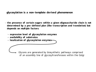
the presence of certain sugars within a given oligosaccharide chain is not determined by a pre-defined plan (like transcription and translation) but depends on multiple factors:. - expression level of glycosylation enzymes - availability of substrates

E N D
the presence of certain sugars within a given oligosaccharide chain is not • determined by a pre-defined plan (like transcription and translation) but • depends on multiple factors: - expression level of glycosylation enzymes - availability of substrates - localization of glycosylation enzymes Glycans are generated by biosynthetic pathways comprised of an assembly line of glycosyltransferases within the Golgi glycosylation is a non-template derived phenomenon
How are the glycosyltransferases and nucleotide-sugar transporters kept in the proper order and in the proper compartment? multiple mechanisms involved - 1) information is contained within the structure or sequence of the enzymes (intrinsic) 2) dynamics of the Golgi (global)
Intrinsic mechanisms: ER retention sequences • the localization of protein O-fucosyltransferase 1 to the ER is mediated by the presence of a KDEL-like sequence in its C-terminal tail • human OFut1: rpssffgmdrppklrdef • fly OFut1: fwgfpkekdrkhtnvheel • (KDEL: classic sequence for receptor-mediated retention of ER resident proteins) • are there short sequences within glycosylation enzymes (i.e. in their cytoplasmic • portions) that mediate Golgi localization and retention? (answer still not clear)
Intrinsic mechanisms: physical and functional associations (part 1) GlcNAcTI was retained in the ER by grafting the cytoplasmic tail of Iip33 to its own tail; endogenous ManII was also retained in the ER suggseting that enzymes interact The two enzymes were later shown to associate via their stem regions (helps ensure that these enzymes act sequentially in N-glycan processing)
pathways of glycolipid biosynthesis Intrinsic mechanisms: physical and functional associations (part 2) GalT, SiaT1, SiaT2 form a trimeric complex Associations between enzymes involved in glycolipid biosynthesis help to shunt substrates along certain pathways and generate specifically glycosylated products
Intrinsic mechanisms: physical and functional associations (part 3) GalCer synthase binds to UDP-galactose transporter and selectively retains a fraction of the total transporter in the ER
IdoA GlcA Intrinsic mechanisms: physical and functional associations (part 4) Association of EXT1 and EXT2 produces a highly active polymerase for HS biosynthesis and is required for Golgi localization Association of GlcA epimerase and 2OST ensures that IdoA is sulfated, limiting its reversion
plasma membrane trans Golgi network (TGN) trans Golgi medial cis ERGIC ER Global mechanisms: Golgi structure and dynamics Two main Golgi functions are: SORTING and GLYCOSYLATION
% of total Golgi proteome What does the Golgi apparatus really look like and what is it composed of? electron micrograph Golgi membranes are composed of several different types of lipid molecules; Golgi proteins can be integral membrane proteins (“resident”) or peripheral proteins that associate with the cytosolic face of the Golgi
UCE and CD-MPR: TGN proteins immunogold electron microscopy How are proteins distributed within Golgi membranes? Although Golgi proteins and enzymes are often concentrated in certain stacks or cisternae, they really exist in a “distribution gradient” within the Golgi and can also be found in other subcellular compartments (i.e. endosomes or ER)
plasma membrane transmembrane domain (Golgi enzyme) transmembrane domain (cell surface receptor) trans Golgi network (TGN) = thickening of the membrane Higher sphingolipid and cholesterol Golgi ER Global/intrinsic mechanisms: lipid bilayer sorting Since Golgi enzymes tend to have shorter transmembrane regions compared to cell surface proteins, it was thought that the thinner bilayer of the Golgi would retain these enzymes and not allow them to reach the plasma membrane
Vesicular transport vs. cisternal maturation: competing models of Golgi dynamics * these two models differ in how secreted proteins move through the Golgi, how Golgi enzymes are retained within the organelle and the nature of the Golgi itself
The vesicular transport model views the Golgi as a stable organelle with secreted proteins being moved between cisternae or stacks in small vesicles glycosylation enzymes are stable“residents” of the Golgi (this model depends heavily on intrinsic mechanisms for Golgi retention)
The cisternal maturation model views the Golgi as a highly dynamic organelle that continuously arises from the ER and is consumed at the trans-Golgi network modes of retrograde transport: - intra-Golgi (enzyme localization) - Golgi-to-ER (lipid balancing) Golgi membranes and glycosylation enzymes are highly mobile (this model depends heavily on retrograde transport and protein/lipid recycling)
What is involved in retrograde transport and vesicle trafficking? • recruitment of proteins into COPI-coated vesicles (many proteins) • budding of vesicles from membranes (ARFs, Rabs, etc.) • transport of vesicles to target membranes (Rab effectors, dynein, kinesin, • microtubules) • tethering of vesicles to target membranes (multiprotein complexes - TRAPP, COG, GARP) • binding and fusion of acceptor and target membranes (SNAREs, SNAP)
An integrated (and therefore complex) view of how both intrinsic and extrinsic mechanisms might contribute to Golgi enzyme localization GREEN - late Golgi enzymes PINK - early Golgi enzymes Early Golgi enzymes move through the stack by cisternal progression, forming large complexes that readily recycle back to early compartments Due to lack of signals or selective partitioning, late Golgi enzymes do not efficiently recycle and thereby concentrated in the TGN
Key Points: • the fidelity of glycan biosynthesis depends, in part, on the order of glycosylation • enzymes within the Golgi apparatus • both structural features of the enzymes and the overall dynamics of the Golgi • contribute to enzyme localization • physical and functional interactions between glycosylation enzymes ensures that • modifications are properly made • the two competing models of Golgi organization, the vesicular transport model and • the cisternal maturation model, differ in howsecreted proteins move through the • Golgi, how Golgi enzymes are retained within the organelle and the nature of the • Golgi itself • an understanding of how Golgi glycosylation enzymes are localized and identification • proteins that regulate this process is slowly emerging
How important are Golgi dynamics and retrograde trafficking in the maintenance of proper glycosylation?