Download
1 / 44
440 likes | 459 Vues
Learn about the basic structure of bone tissue, different types of bone, bone marrow, bone vessels, and the anatomy of the vertebral column and ribs.

E N D
Introduction to osteoloy Skeleton of the spine and thorax
the basic structure of the bone tissue • bone isconnectivetissue, white, tough but flexible • Itdevelops as activityofosteoblasts, whichproduceprimary bone massand thentheychange in theosteocytes • a) osteocytes – located in cavities(lacuna) withintheprimary bone mass • b) Primary bone mass– containsorganiccomponent (ossein) and anorganiccomponent (calciumphosphate)
We know two forms of bone tissue a) Compact bone (substantia compacta) b) Spongy bone (substantia spongiosa)
Compact bone H – osteon 1 – osteocyte 2 – lacuna 3 – canaliculus osseus 4 – Haversian canal 1 – Haversovs lamells 2 – intersected lamells 3 – superficial lamells 4 – spongy bone
Substantia spongiosa • Substantia compacta • Skull - diploe
PERIOSTEUM a) Fibrous layer(externaů) b) Cambious layer (internal) – rich sensory innervation 1– periost 2 – Sharpey fibres 3 – vessels 4 – endost
BONE MARROW Medulla ossium rubra – red bone marrow (active hematopoetic tissue) Medulla ossium flava – yellow bone marrow Medulla ossium gelatinosa – grey bone marrow
Long bones Middle section (diaphysis) Ends (epiphysis) proximal and distal Medullar cavity
Short bones (various shapes) 1) On the surface - corticalis 2) Inside - substantia spongiosa
Flat bones • Compact bone has twolayers: • lamina externa and interna and betweenthem, • thereisspongy bone – diploe
Sesamoidal bones In some muscle ligaments Pneumatized bones
BONE VESSELS • The most importatnt bone vessels come through periosteum via Volkmann channels
Blood supply of the long bone aa. epiphysariae aa. metaphysariae a. nutricia Periostal vessels
X-RAY PICTURES 4,5 years 7years 11 years 14 years
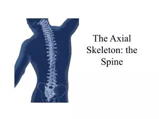
COLUMNA VERTEBRALIS - SPINE During development: 33-34 vertebrae After fusion: 24 vertebrae Vertebrae 7 cervical 12 thoracic 5 lumbar 4-5 sacral- os sacrum 4-5 coccygeal- os coccygis
VERTEBRA corpus vertebrae facies terminalis superior et inferior arcus vertebrae pediculus arcus vertebrae lamina arcus vertebrae foramen vertebrale incisura vertebralis processus processus articulares processus transversi processus spinosus
CERVICAL VERTEBRAE uncus corporis vertebrae processus transversus -tubercula anteriora et posteriora, foramina processus transversi oval body Triangular foramen vertebrale cleft processus spinosus processus articulares – in oblique plane
Corpus vertebrae Arcus vertebrae Processus articulares Processus transversus Processus spinosus Costa
ATLAS - C1 arcus anterior tuberculum anterior fovea dentis arcus posterior tuberculum posterior sulcus a. vertebralis massae laterales processus transversi foramina pr. transversi
Corpus vertebrae Arcus vertebrae Processus articulares Processus transversus Processus spinosus Costa
AXIS - C2 dens axis (original body of atlas)- apex dentis facies articularis anterior et posterior os odontoideum
Corpus vertebrae Arcus vertebrae Processus articulares Processus transversus Processus spinosus Costa
C6- TUBERCULUM CAROTICUM VERTEBRA PROMINENS- C7
VERTEBRAE THORACICAE • corpus: foveae costales - superiores, inferiores • processus transversus 1.-10.Th: fovea costalis pr. transversi • processus articulares: in frontal plane • Th4 – 9: impressio aortica
Corpus vertebrae Arcus vertebrae Processus articulares Processus transversus Processus spinosus Costa
VERTEBRAE LUMBALES • processus costarii • processus accesorius- more caudally • processus mamillaris- more cranially • processus articulares- in sagital plane • processus spinosi- flat plate • Sacralization of last lumbar vertebra
Corpus vertebrae Arcus vertebrae Processus articulares Processus transversus Processus spinosus Costa
OS SACRUM • facies dorsalis- crista- mediana, medialis, lateralis • facies auricularis- partes laterales ossis sacri • facies pelvina- lineae transversae • foramina sacralia- dorsalia, pelvina • canalis sacralis- hiatus sacralis- cornua sacralia • basis ossis sacri • apex ossis sacri • Lumbalization of sacral vertebra
Corpus vertebrae Arcus vertebrae Processus articulares Processus transversus Processus spinosus Costa
OS COCCYGIS • cornua ossis coccygis= processus transversi Co1 • apex coccygis Corpus vertebrae Processus articulares Processus transversus
VÝVOJ OBRATLŮ Corpus vertebrae Arcus vertebrae Processus articulares Processus transversus Processus spinosus Costa
COSTAE - RIBS • 12 pairs of ribs: • costae verae: 7 pairs, true ribs • costae spuriae: 8th-10th pair, false ribs • costae fluctuantes (liberae) : 11th and 12th pair- free ribs • length- from 1st to 8th increases, the smallest: 1st anf 12th, the largest 6th – 9th
RIB os costae + cartilago: caput costae, crista collum costae tuberculum costae corpus costae crista costae sulcus costae angulus costae
Facies articularis capitis costae Facies articularis tuberculi costae
COSTA PRIMA • sulcus arterie subclaviae • (sulcus venae subclaviae) • tuberculum m. scaleni anterioris • úpon pro m. scalenus medius • - odstup m. subclavius
COSTA SECUNDA/second rib - tuberculum m. scaleni posterioris - tuberositas m. serrati anterioris
STERNUM • sternebrae • manubrium sterni- incisura- jugularis, clavicularis and places for connection with cartilages of the first pair of ribs • angulus sterni • corpus- incisurae costales • processus xiphoideus
Thank you for your attention! Obrázky: Atlas der Anatomie des Menschen/Sobotta. Putz,R., und Pabst,R. 20. Auflage. München:Urban & Schwarzenberg, 1993 Netter: Interactive Atlas of Human Anatomy. Naňka, Elišková: Přehled anatomie. Galén, Praha 2009. Čihák: Anatomie I, II, III. Drake et al: Gray´s Anatomy for Students. 2010