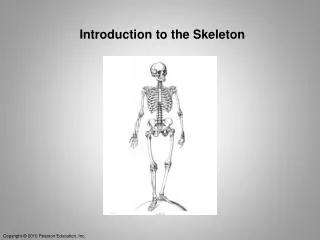
Introduction to the Skeleton
Introduction to the Skeleton. The human skeleton is made of both cartilage and bone tissue. An infant’s skeleton is primarily cartilage but as the infants grows it is replaced by bone. Bones and Skeletal Tissue. Figure 6.17 Fetal primary ossification centers at 12 weeks. Parietal bone.

Introduction to the Skeleton
E N D
Presentation Transcript
The human skeleton is made of both cartilage and bone tissue. An infant’s skeleton is primarily cartilage but as the infants grows it is replaced by bone. Bones and Skeletal Tissue
Figure 6.17 Fetal primary ossification centers at 12 weeks. Parietal bone Occipital bone Frontal bone of skull Mandible Clavicle Scapula Radius Ulna Ribs Humerus Vertebra Ilium Tibia Femur
Cartilage consists primarily of water. This provides resilience and compressibility that protects the bones. There are three types: Skeletal Cartilage
Cartilage consists primarily of water. This provides resilience and compressibility that protects the bones. There are three types: Hyaline Elastic Fibrocartilage Skeletal Cartilage
Hyaline Cartilage • Most abundant • Chondrocytes appear spherical • Collagen is the only fiber Locations: • Articular cartilage • Costal cartilage • Respiratory cartilage • Nasal cartilage
Fibro elastic Cartilage • Very compressible, chondrocytes are found in parallel rows • Found only in vertebral discs and meniscus of the knee.
Elastic Cartilage • Similar to hyaline but contains elastic fibers • Found in areas where there is repeated bending • Two locations: • External ear • Epiglottis
Figure 6.1 The bones and cartilages of the human skeleton. Epiglottis Larynx Thyroid cartilage Cartilage in external ear Cartilages in nose Trachea Cricoid cartilage Lung Articular Cartilage of a joint Cartilage in Intervertebraldisc Costal cartilage Respiratory tube cartilages in neck and thorax Bones of skeleton Pubic symphysis Axial skeleton Meniscus (padlike cartilage in knee joint) Appendicular skeleton Cartilages Articular cartilage of a joint Hyaline cartilages Elastic cartilages Fibrocartilages
Perichondrium This supplies nutrients via diffusion to the avascular cartilage. This limits the thickness of the cartilage.
Chondrocytes The chondrocyte is the cell responsible for the maintenance and growth of the cartilage. They reside in lacunae
Cartilage can grow two ways: Appositional growth Interstitial growth Cartilage Growth
Appositional Growth Appositional growth (from the edges) occurs where cells from the perichondrium lay down new a cartilage matrix.
Interstitial Growth Growth from inside the cartilage. The chondrocytes divide in the lacunae laying down more matrix.
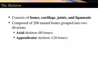
There are 206 bones in the adult skeleton. Skeletal Classification
The skeleton is divided into the: Axial Skeleton Appendicular Skeleton Skeletal Classification
Axial Skeleton Forms the long axis of the body. Its primary roll is support and protection of the organs.
Appendicular Skeleton Bones of the upper and lower limbs including the shoulder and hips. It is involved in locomotion.
Bones are further classified by shape. There are 4 types: Long Bones Short Bones Flat Bones Irregular Bones Bone Classification
Long Bones Are longer than wide. It consists of a shaft plus two ends.
Long Bones Are longer than wide. It consists of a shaft plus two ends. Found on the Appendicular Skeleton.
Long Bones Are longer than wide. It consists of a shaft plus two ends. Found on the Appendicular Skeleton. Size doesn’t matter Femur vs phalanges
Short Bones Typically cube shape. These are found on the wrist (carpals) and ankle (tarsals)
Flat Bones Are flat and thin in 1 dimension. Often have a curve edge Examples include ribs, scapula and skull
Irregular Shape No specific shape, examples include the pelvic bones and vertebrae.
Figure 6.2 Classification of bones on the basis of shape. (c) Flat bone (sternum) (a) Long bone (humerus) (b) Irregular bone (vertebra), right lateral view (d) Short bone (talus)
Support- respiration Protection-skull Movement-Appendicular skeleton Mineral Storage-calcium phosphate Blood Cell Formation- Hematopoiesis Fat Storage Function of the Skeletal System
Bone is can be classified as compact or spongy bone. Gross anatomy of Bone
Gross anatomy of Bone Compact bone is dense and has a smooth outer surface.
Gross anatomy of Bone Spongy bone has a honey comb structure made up of small projections called trabeculae.
Diaphysis is the long central shaft of a long bone Structure of the long bone
Diaphysis is the long central shaft of a long bone Epiphyses are the ends of the bone Structure of the long bone
Diaphysis is the long central shaft of a long bone Epiphyses are the ends of the bone Membranes cover the exterior and interior of the bones Structure of the long bone
Diaphysis is the long central shaft of a long bone Epiphyses are the ends of the bone Membranes cover the exterior and interior of the bones Composed both of compact and spongy bone Structure of the long bone
Structure of the long bone • The diaphysis has a large central cavity and is filled with fat (yellow marrow).
Structure of the long bone • The epiphyses are filled with hematopoietic tissue (red marrow) which gives rise to the blood cells.
The periosteum covers the exterior of the bone. It contains bone forming cells and is involved with bone growing wider. Structure of the long bone
Articular cartilage Proximal epiphysis Spongy bone Epiphyseal line Periosteum Compact bone Medullary cavity (lined by endosteum) Diaphysis Distal epiphysis (a)
Figure 6.3c The structure of a long bone (humerus of arm). Endosteum Yellow bone marrow Compact bone Periosteum Perforating (Sharpey’s) fibers Nutrient arteries (c)
Four cell types populate the bone tissues. These are: Osteogenic cells Osteoblasts Osteocytes Osteoclasts Microscopic Anatomy of Bone
Osteogenic cells are stem cells that give rise to other bone forming cells Microscopic Anatomy of Bone
Osteoblasts are bone forming cells Osteocytes maintains the bone matrix Osteoclasts destroy bone Microscopic Anatomy of Bone
Figure 6.4 Comparison of different types of bone cells. (a) Osteogenic cell (b) Osteoblast (c) Osteocyte (d) Osteoclast Stem cell Matrix-synthesizing cell responsible for bone growth Mature bone cell that maintains the bone matrix Bone-resorbing cell
The structural unit of the compact bone is the osteon or Haversian System. It consists of a central canal that contains blood vessels and nerves. Microscopic Anatomy Compact Bone
Branches from the nerves and blood vessels go transversely through Volkmann's canals. Microscopic Anatomy Compact Bone
Figure 6.7 Microscopic anatomy of compact bone. Spongy bone Compact bone Central (Haversian) canal Perforating (Volkmann’s) canal Endosteum lining bony canals and covering trabeculae Osteon (Haversian system) Circumferential lamellae (a) Perforating (Sharpey’s) fibers Periosteal blood vessel Lamellae Periosteum Nerve Vein Lamellae Artery Central canal Lacuna (with osteocyte) Canaliculi Osteocyte in a lacuna Lacunae Interstitial lamellae (b) (c)
Microscopic Anatomy Compact Bone Osteocytes are found in the lacunae.
Bone has an inorganic and organic component. The inorganic component is calcium phosphate. The organic component is made up of collagen and proteoglycans. Bone Formation (Remodeling)
Bone is unique because is can repair itself and adjust to mechanical stresses. Bone Formation (Remodeling)
Bone is unique because is can repair itself and adjust to mechanical stresses. This is a complex process involving a tug of war between Osteoclasts and Osteoblasts. Bone Formation (Remodeling)