Anatomy of the Human Skeleton: Structure and Function
Learn about the composition, functions, and regions of the human skeleton, including the skull and axial skeleton. Understand the bone structure, joints, ligaments, and cavities of the skull. Discover the intricate details of facial and cranial bones.

Anatomy of the Human Skeleton: Structure and Function
E N D
Presentation Transcript
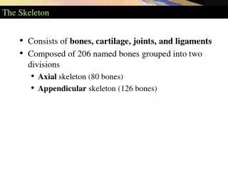
THE SKELETON • Composed of bones, cartilages, joints, and ligaments, accounts for about 20% of body mass (30 pounds in a 160 pound person) • Bones make up most of the skeleton • Cartilages occur only in isolated areas • Nose, parts of the ribs, and the joints • Ligamentsconnect bones and reinforce joints, allowing required movements while restricting motions in other directions • Joints, the junction between bones, provide for the remarkable mobility of the skeleton
Axial Skeleton • Structured from 80 bones segregated into three major regions: • Skull • Vertebral column • Bony thorax • Supports the head, neck, and trunk, and protects the brain, spinal cord, and organs in the thorax
The Skull • The skull consists of22 cranial and facial bones that form the framework of the face, contain cavities for special sense organs, provide openings for air and food passage, secure the teeth, and anchor muscles of facial expression • The cranial bones (cranium) enclose and protect the fragile brain and furnish a site for attachment of head and neck muscles • Facial bones: • Form the framework of the face • Contain cavities for the special sense organs of sight, taste, and smell • Provide openings for air and food passage • Secure the teeth • Anchor the facial muscles of expression, which we use to show our feelings
The Skull • Except for the mandible, which is joined to the skull by a freely movable joint, most skull bones are flat bones joined by interlocking joints called sutures • Suture lines have a saw-toothed or serrated appearance • Major sutures that connect cranial bones are: • Coronal • Sagittal • Squamous • Lambdoid
The Skull • Most other skull sutures connect facial bones and are named according to the specific bones they connect • Examples: • Frontonasal suture • Occipitomastoid suture
Overview of Skull Geography • The anterior aspect of the skull is formed by facial bones, and the remainder is formed by a cranium, which is divided into: • 1. Cranial vault (calvaria): • Forms the superior, lateral, and posterior aspects of the skull, as well as the forehead
The Skull • 2. Cranial base(floor) (a): inferior superficial view • Forms the skull’s inferior aspect • Internally (b+c): • (b): superior view of the floor of cranial cavity • (c): schematic view of the cranial cavity floor • prominent bony ridges divide the base into three distinct “steps” or fossa: • Anterior cranial fossa • Middle cranial fossa • Posterior cranial fossa
The Skull • The cavities of the skull include: • Cranial cavity: • Houses the brain • Ear cavities • Nasal cavity • Orbit cavities: • House the eyeballs • Air-filled sinuses: • Lighten the skull
The Skull • The skull has about 85 named openings that provide passageways for the spinal cord, major blood vessels serving the brain, and the cranial nerves • Named: • Foramina • Canals • Fissures
Cranium • Consists of eight strong, superiorly curved bones • 1. Frontal bone: • Forms the anterior cranium • Most anterior part of the frontal bone is the vertical frontal squama (forehead) • Articulates posteriorly with the parietal bones via the coronal suture, extends forward to the supraorbital margins, and extends posteriorly to form the superior wall of the orbits and most of the anterior cranial fossa
Parietal Bone • 2. Two large, rectangular bones on the superior and lateral aspects of the skull • Form the bulk of the cranial vault • The four largest sutures of the skull are located where the parietal bones articulate with other bones: • Coronal: parietal bones meet the frontal anteriorly • Sagittal: the parietal bones meet superiorly at the cranial midline • Lambdoid: parietal bones meet the occipital bone posteriorly • Squamous: where a parietal and temporal bone meet on the lateral aspect of the skull
Occipital Bone • Articulates with the parietal, temporal, and sphenoid bones • Forms most of the posterior wall and base of the skull • The foramen magnum, a large opening through which the brain connects to the spinal cord, is located in the base of the occipital bone • Rockerlike occipital condylesarticulate with the first vertebra of the spinal column in a way that permits a nodding movement of the head
Temporal Bone • Lateral skull surface • Articulate with the parietal bones and form the inferolateral aspects of the skull and parts of the cranial floor • The temporal bone is characterized by the small, oval mandibular fossa on the inferior surface of the zygomatic process • It receives the condyle of the mandible (lower jawbone) forming the freely movable temporomandibular joint
Temporal Bone • Tympanic region surrounds the externalacoustic meatus (external ear canal) • Housed in the petrous region are the middle and inner ear cavities, which contain sensory receptors for hearing and balance
Temporal Bone • Jugular foramen: passage of internal jugular vein and three cranial nerves • Carotid canal: passage of the internal carotid arteries (both supply 80% of blood to the brain) • Closeness to the inner ear cavities explains why, during excitement or exertion, we sometimes hear our rapid pulse as a thundering sound in the head • Mastoid process: felt as a bump just posterior to the ear • Anchoring site for some neck muscles • Full of air cavities (mastoid sinuses: air cells) • Position adjacent to the middle ear cavity (high-risk area for infections spreading from the throat) puts it at risk for infection itself • Mastoid sinus infection (mastoiditis) is difficult to treat • Separated from the brain by only a very thin bony plate • Infections may spread to the brain
Sphenoid Bone • Spans the width of the middle cranial fossa (furrow or shallow depression) • Keystone of the cranium because it articulates with all other cranial bones • Pterygoid processes anchor the pterygoid muscles which are important in chewing • Optic canals: allow passage of optic nerve
Ethmoid Bone • Lies between the sphenoid and nasal bones • Forms most of the bony area between the nasal cavity and the orbits • Superior surface (cribriform plate) helps form the roof of the nasal cavities and the floor of the anterior cranial fossa • Olfactory foramina: allows passage of the olfactory nerve • Perpendicular plate: forms superior part of the nasal septum, which divides the nasal cavity into right and left halves
Sutural Bones • Sutural, or Wormian, bones are groups of irregularly shaped bones or bone clusters located within sutures (most often in the lambdoid suture) that vary in number and are not present on all skulls • Formed during fetal development • Structurally unimportant
Facial BonesMandible • U-shaped bone • Lower jawbone • Largest, strongest bone of the face • Body forms the chin • Two upright rami • Each ramus meets the body posteriorly at a mandibular angle • Superior margin of each ramus are two processes separated by the mandibular notch • The anterior coronoid process is an insertion point for the large temporalis muscle that elevates the lower jaw during chewing • The posterior mandibular condyle articulates with the mandibular fossa of the temporal bone, forming the temporomandibular joint on the same side
Facial BonesMandible • Mandibular body anchors the lower teeth: • Its superior border, called the alveolar margin, contains the sockets (alveoli) in which the teeth are embedded • In the midline of the mandibular body is a slight depression, the mandibular symphysis, indicating where the two mandibular bones fused during infancy