The Axial Skeleton
800 likes | 873 Vues
Explore the anatomy of the cranium and facial bones, learn about skull divisions, fossae, openings, and sutures. Dive into the details of cranial bones, their functions, and the anatomy of the skull. Discover the significance of the brain's placement in the cranial fossae.

The Axial Skeleton
E N D
Presentation Transcript
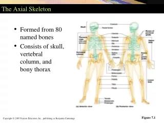
7 The Axial Skeleton
Repition….Repition….Repition…. Repition….Repition….
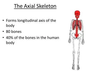
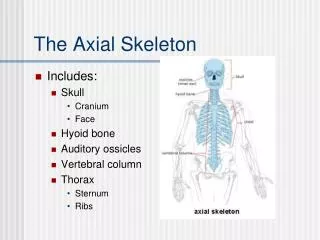
I. The Skeleton A. Bones, cartilage, joints, and ligaments 1. joints - also called articulations B. 206 named bones grouped into two divisions 1. Axial skeleton (80 bones) ► skull, vertebral column, and thoracic cage 2. Appendicular skeleton (126 bones) ► upper and lower limbs
II. The Cranium (Skull) A. The cranium is formed by cranial and facial bones 1. enclose and protect the brain 2. provide attachment sites for muscles of the head and neck B. Facial bones 1. Form framework of the face 2. Form cavities for sense organs of sight, taste, and smell 3. Provide openings for passage of air and food 4. Hold the teeth in place 5. Anchor muscles of the face
C. Internally bony ridges divide skull into distinct fossae (surfaces) 1. anterior 2. middle 3. posterior D. Brain sits within the cranial fossae 1.brain occupies cranial cavity
Anterior cranialfossa Middle cranialfossa Posterior cranialfossa Superior view of the cranial fossae
E. The skull contains smaller cavities 1. middle & inner ear cavities—in lateral aspect of cranial base 2. nasal cavity—lies in and posterior to the nose 3. orbits—house the eyeballs 4. sinuses — air-filled spaces in bones around the nasal cavity
F. The skull contains approximately 85 named openings 1. foramina, canals, and fissures 2. provide openings for important structures a. spinal cord b. blood vessels serving the brain c. 12 pairs of cranial nerves
IV. Cranial Bones A. Formed from eight large bones 1. paired bones a. temporal bones b. parietal bones 2. unpaired bones a. frontal bone b. occipital bone c. sphenoid bone d. ethmoid bone
V. Sutures A. Four sutures of the cranium 1. coronal suture - runs in the coronal plane 2. squamous suture - parietal bone meets temporal bone 3. sagittal suture - where right and left parietal bones 4. lambdoid suture – parietal bones meet the occipital bone
right side of the skull Frontal bone Coronal suture Sphenoid bone Parietal bone Squamous suture Ethmoid bone Lacrimal bone Lambdoid suture Lacrimal fossa Occipital bone Nasal bone Temporal bone Zygomatic process Zygomatic bone Maxilla External acoustic meatus Mastoid process Styloid process Mandible Condylar process Mental foramen Mandibular notch Coronoid process Mandibular ramus Mandibularangle
Frontal bone Parietal bone Supraorbital foramen Nasal bone Sphenoid bone Superior orbital fissure Temporal bone Optic canal Ethmoid bone Inferior orbital fissure Lacrimal bone Zygomatic bone Infraorbital foramen Maxilla Vomer Mandible Mentalforamen
Parietal bone Sagittal suture Lambdoidsuture Occipital bone Superior nuchal line External occipitalprotuberance Inferior nuchal line Occipital condyle
Maxilla Hardpalate Palatine bone Infraorbital foramen Maxilla Sphenoid bone Zygomatic bone Vomer zygomatic process External acoustic meatus Styloid process Mastoid process Jugular foramen Temporal bone Occipital condyle Occipital bone External occipitalprotuberance Foramen magnum Inferior view of the skull (mandible removed)
Externalacoustic meatus Zygomaticprocess Mastoid process Styloidprocess
Optic canal Lesser wing Foramenrotundum Greater wing Foramen ovale Sellaturcica Foramen spinosum Body of sphenoid Superior view
Crista galli Cribriform platewith cribriformforamina
The Holes in the Head Optic foramen Foramen rotundum Foramen ovale Foramen spinosum Foramen lacerum Internal acoustic meatus Jugular foramen Hypoglossal canal Foramen magnum ▲
Crista galli Ethmoidbone View Cribriform plate Frontal bone Optic canal Lesser wing Sphenoid Greater wing Foramen rotundum Foramen ovale sella turcica Foramen spinosum Foramen lacerum Internal acousticmeatus Temporal bone Jugular foramen Hypoglossal canal Parietal bone Occipital bone Foramen magnum Superior view of the skull, calvaria removed
View Superior view of the skull, calvaria removed
The Sphenoid Bone Optic canal Lesser wing Foramenrotundum Greater wing Foramen ovale Sellaturcica Foramen spinosum Body of sphenoid Superior view
Parietal bone Coronal suture Frontal bone Sphenoid bone Squamoussuture Temporal bone Crista galli Lambdoid suture Nasal bone Occipital bone Ethmoid bone Vomer External occipitalprotuberance Maxilla Internal acousticmeatus Mandible Mandibularforamen Palatine bone Midsagittal section showing the internal anatomy of the left half of skull
Midsagittal section showing the internal anatomy of the left half of skull
Mandible Temporomandibularjoint Coronoidprocess Mandibular notch Condylarprocess Mandibular foramen Ramusofmandible Mental foramen Mandibularangle Body of mandible Mandible, right lateral view
Mandible Mandible, right lateral view
Figure 7-15a Paranasal sinuses. Frontalsinus Ethmoidalair cells(sinus) Sphenoidalsinus Maxillarysinus Anterior aspect
Figure 7.17 The hyoid bone. Greater horn Lesser horn Body
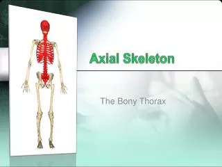
XIII. Thoracic Cage Jugular notch Clavicular notch Manubrium Sternal angle Body Sternum Trueribs(17) Xiphoidprocess Falseribs(812) Costalcartilage L1Vertebra Floatingribs (11, 12)
XIII. Thoracic Cage L1Vertebra
The Vertebral Column A. In the adult, is formed from 26 bones B. Transmits weight of trunk to the lower limbs C. Surrounds and protects the spinal cord
Regions and Normal Curvatures A. The vertebral column has five major regions ► 7 cervical vertebrae of the neck region ► 12 thoracic vertebrae ► 5 lumbar vertebrae ► Sacrum - five fused bones ► Coccyx - inferior to sacrum
C1 2 3 (concave) Cervical curvature 7 vertebrae, C1 C7 4 5 6 7 T1 Spinousprocess 2 3 Transverseprocesses 4 5 Thoracic curvature 6 (convex) 12 vertebrae, T1 T12 7 8 9 Intervertebraldiscs 10 Intervertebralforamen 11 12 L1 2 Lumbar curvature (concave) 5 vertebrae, L1 L5 3 4 5 Sacral curvature (convex) 5 fused vertebraesacrum Coccyx 4 fused vertebrae Right lateral view Anterior view
Regions and Normal Curvatures A. Curvatures of the spine 1. Cervical and lumbar curvatures ► Concave posteriorly 2. Thoracic and sacral curvatures ► Convex posteriorly kyphosis – exagerated curvature in thoracic region (humpback) lordosis – exaggerated curvature in the lumbar region scoliosis – S-shaped curvature of the whole vertebral column
Posterior longitudinalligament Anterior longitudinalligament Body of a vertebra Intervertebral disc Anterior view of part of the spinal column
Intervertebral Discs A. Are cushion-like pads between vertebrae 1. Nucleus pulposus a. gelatinous inner sphere b. absorbs compressive stresses 2. Anulus fibrosus a. outer rings formed of ligament b. inner rings formed of fibrocartilage c. contains the nucleus pulposus
Posterior Vertebralarch Lamina Spinousprocess Transverseprocess Superiorarticularprocessandfacet Vertebralforamen Pedicle Body Anterior
Posterior Anterior
Cervical Vertebrae A. Cervical 1. C1 (atlas) no body, no spine 2. C2 (axis) bifid spine, dens (head) 3. C3-6 bifid spine 4. C7 non-bifid spine, first bulge in lower neck 5. transverse foramen (vessel+nerve) 6. bodies get larger in descending fashion
Dens of axis Transverse ligamentof atlas C1 (atlas) C2 (axis) C3 Inferior articular process Bifid spinous process Transverse processes C7 (vertebra prominens) Cervical vertebrae