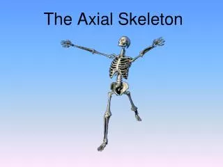
Overview of the Axial Skeleton: Head and Trunk Framework
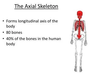
This lecture delves into the axial skeleton, composed of 80 bones that form the bony framework of the head and trunk. It covers the cranium with its eight distinct cranial bones, including the frontal, parietal, temporal, ethmoid, sphenoid, and occipital bones, each with unique functions and features such as sinuses and attachment points. The discussion extends to the vertebral column, comprising 26 vertebrae in adults, and includes details about the thorax's components, including the sternum and ribs, highlighting their structural significance and connectivity.

Overview of the Axial Skeleton: Head and Trunk Framework
E N D
Presentation Transcript
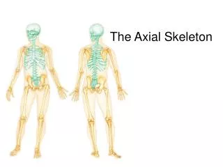
The Axial Skeleton Divisions of the Skeleton Lecture 2
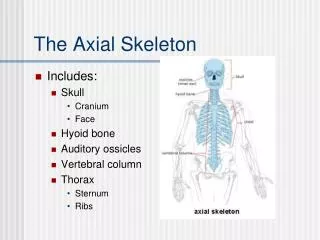
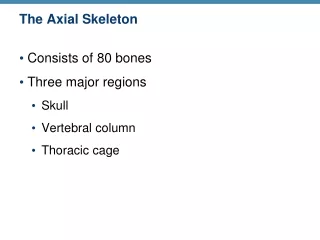
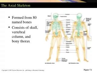
Bony Framework of the Head and Trunk • Consists of 80 bones • Framework of the Skull • Cranium • Composed of eight distinct cranial bones • Frontal bone: forms the forehead • Frontal sinuses communicate with the nasal cavities • Sinuses near the nose are described as paranasalsinuses • Parietal bones: form most of the top and side walls
Temporal bones: form part of the sides and some of the base • Each contains a mastoid sinus as well as the ear canal, the eardrum, and the entire middle and internal ears. • The mastoid process contains the mastoid air cells and serves as a place for muscle attachment. • Ethmoid bone: located between the eyes • Contains several air spaces, comprising some of the paranasal sinuses
Sphenoid bone: forms part of the eye socket • It contains a saddle-like depression, the sellaturcica, that holds and protects the pituitary gland • Occipital bone: forms the back and part of the base of the skull • The foramen magnum is a large opening through which the spinal cord communicates with the brain.
Skull Frontal Parietal Temporal Zygomatic Maxilla Occipital Mandible
Facial Bones • Fourteen bones that make up the face • Mandible: a.k.a. the lower jaw bone, is the only movable bone of the skull • Maxillae bones: fuse at the midline to form the upper jaw bone • Each maxilla contains a large air space, the maxillary sinus, that communicates with the nasal cavity. • Zygomatic bones: form the prominences of the cheeks • Nasal bones: lie side by side, forming the bridge of the nose
Lacrimal bones: lie near the inside corner of the eye in the front part of the medial wall of the orbital cavity • Vomer bones: forms the lower part of the nasal septum • Palatine bones: form the back of the hard palate • Inferior nasal conchae: extend horizontally along the lateral wall of the nasal cavities • Hyoid bone: found just below the skull proper to which the tongue and other muscles are attached
Framework of the Trunk: Vertebral Column • Is made of a series of irregularly shaped bones. • There are 33 or 34 in a child, but fusion reduces this number to 26 separate bones in an adult. • Vertebrae have a drum-shaped body, the centrum, located anteriorly that serves as the weight-bearing part; disks of cartilage between the vertebrae act as shock absorbers and provide flexibility. • A foramen is found in the center of each vertebra, and when ligaments link all of the vertebrae, the spinal canal is formed.
The spinal processes are located posteriorly and can usually be felt just beneath the skin of the back. • Types of vertebrae • Cervical vertebrae: seven in number, located in the neck • The first is the atlas; it supports the head • The second is the axis; it serves as a pivot for the head • Thoracic vertebrae: twelve in number, located in the chest • The posterior ends of the twelve pair of ribs attach to these vertebrae
Lumbar vertebrae: five in number, located in the lower back • Larger and heavier that the other vertebrae to support more weight • Sacral vertebrae: five separate bones in a child; they fuse to form the sacrum in an adult • Coccyx: consists of four or five tiny bones in a childe; they fuse to form a single bone in an adult
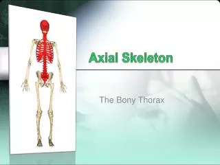
Bones of the Thorax • Sternum: breast bone • Manubrium: the top portion • It joins at the top on each side with the clavicle and joins laterally with the first pair of ribs • Sternal angle: a slight elevation where the manubrium joins the body of the sternum • Xiphoid process: the lower end of the sternum; is cartilaginous in youth and ossifies in adults
Ribs • True ribs: first seven pairs; attached directly to the sternum by individual extensions called costal cartilage • False ribs: remaining five pair; the 8th, 9th, and 10th pairs are attached to the cartilage of the rib above. The last two have no anterior attachments and are known as floating ribs. • Intercostal spaces: the spaces between the ribs containing muscles, blood vessels, and nerves.